Diabetic Macular Edema
Clinical Features:
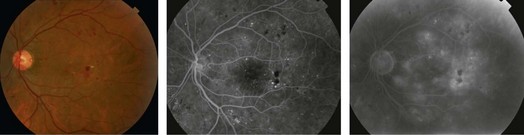
The classic clinical descriptions of DME includes focal, diffuse and cystoid (CME), based on the clinical and angiographic appearance (Fig. 13.2.1). Focal macular edema is characterized by focal leaking microaneurysms giving a well-circumscribed area of thickening often associated with hard exudates. Diffuse macular edema is characterized by more widespread vascular abnormalities giving larger areas of thickening, a paucity of hard exudates and cystic changes in the retina. CME associated with DME appears similar to CME from other causes. It is not unusual for affected eyes to manifest two or all three of these sub-types.
OCT Features (Figs 13.2.2 and 13.2.3):
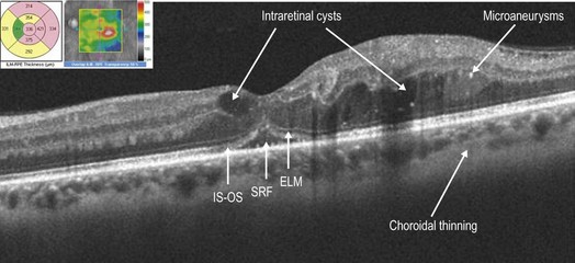
Figure 13.2.2 OCT scanning through the retina shows thickening with outer retinal cystic changes (arrows). The area and extent of thickening can be followed by the false color rendering of the thickness map over the C-scan (inset). The retinal thickness map also provides quantitative information about thickening and is useful in gauging effect of treatment. Also note the hyper-reflective clusters in the outer retina, the trace subretinal fluid or SRF (arrow) and that the external limiting membrane is relatively well-preserved in this patient (arrow) while the IS–OS/ellipsoid layer shows some disruption centrally (arrow).
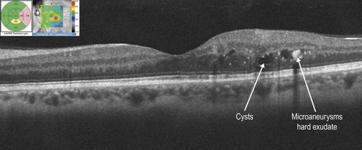
Figure 13.2.3 OCT scan of the same patient after focal laser therapy. The edema and cysts are reduced, as is the retinal thickness on the thickness map. Also, the normal architecture of the IS–OS ellipsoid layer seems relatively well restored.
The OCT appearance of DME can be categorized into four major types:
▶ Thickening of the fovea with homogenous optical reflectivity throughout the whole layer of the retina.
▶ Thickening of the fovea with markedly decreased optical reflectivity in mostly the outer retinal layers (cystoid changes).
▶ Thickening of the fovea with subfoveal fluid accumulation and distinct outer border of detached retina.
▶ Thickening of the fovea with epiretinal membrane formation with or without apparent vitreofoveal traction.
The qualitative assessment of OCT scans in DME are proving increasingly important in predicting outcome as well as determining which patients will respond best to individualized treatment. Moreover, OCT may also show foveal microstructural changes such as disruption of the IS–OS/ellipsoid layer and of the external limiting membrane, which may be correlated to visual acuity in DME. Presence of hyper-reflective foci may also be associated with severity of the edema in DME and may reduce significantly with successful treatment of edema.