Intrauterine Device Location, Abnormal

Published on 10/03/2015 by admin

Filed under Obstetrics & Gynecology

Last modified 22/04/2025

Print this page

rate 1 star rate 2 star rate 3 star rate 4 star rate 5 star
Your rating: none, Average: 0 (0 votes)

This article have been viewed 1563 times

Intrauterine Device Location, Abnormal

Synonyms/Description

Mirena
ParaGard
IUD
IUCD (intrauterine contraceptive device)
LARC (long-acting reversible contraception)

Etiology

The uterus may be too small to accommodate an intrauterine device (IUD) or the IUD may not open normally because of placement or anatomic abnormalities or it may become lodged in the lower uterine segment/cervix. The IUD may become embedded in the myometrium.

Ultrasound Findings

Three-dimensional ultrasound is crucial to determining the location of an IUD. Although the shaft of the IUD is visible using 2-D ultrasound, it is often more difficult to see the relationship between the arms of the IUD and the uterine cavity, especially if the IUD did not open normally or has become bent. Sometimes the shadow of the IUD is much easier to see than the IUD itself, especially when dealing with the non–copper–containing type. The shadow of the IUD often facilitates finding the IUD by following the shadow sonographically.

Differential Diagnosis

If an IUD is in the uterus, there is most often no other diagnosis to consider. If there is a shadowing structure in the uterus, however, other etiologies can include an area of calcification from a fibroid or endometrial scarring, or a foreign body such as a laminaria. If a patient has an ultrasound immediately after an endometrial biopsy or other instrumentation of the cavity, air left in the cavity can be very bright and echogenic with a shadow, simulating an IUD. Generally, the IUDs are a typical shape that is recognizable and not usually confused with these other conditions.

Clinical Aspects and Recommendations
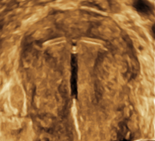
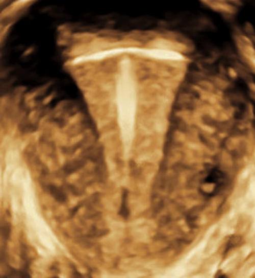
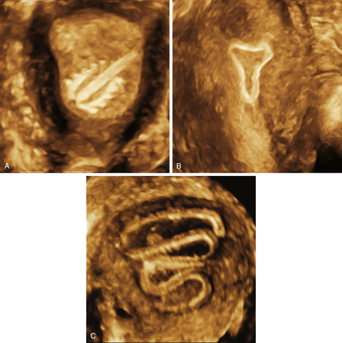
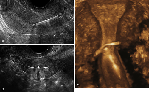
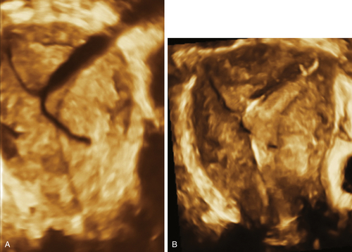
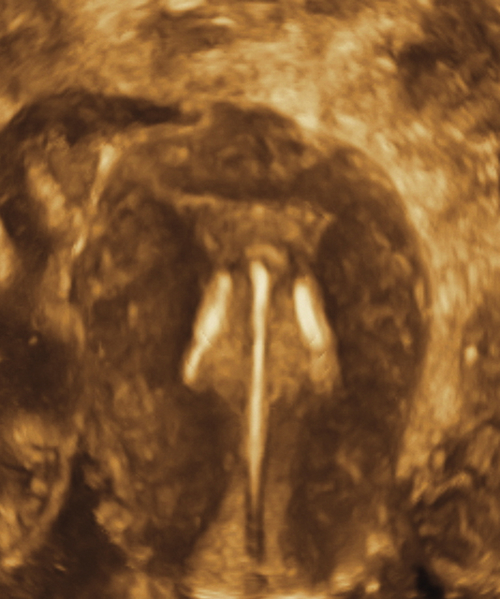
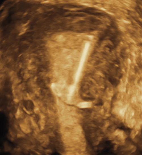
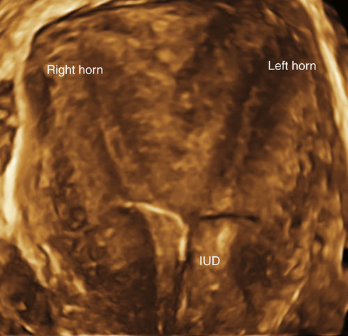
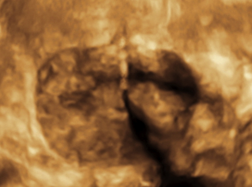
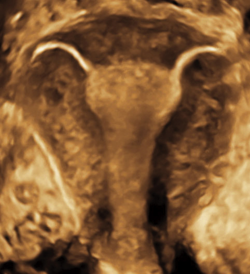
An abnormally located IUD can cause pelvic pain and bleeding, although this can also be an incidental finding in an asymptomatic patient. If a patient with an IUD presents with pain or bleeding, a 3-D ultrasound should be done to evaluate the position of the device in the uterus. Malpositioned IUDs need to be removed to improve symptoms.

Figures

image

Figure I1-1 Mirena IUD in place.

image

Figure I1-2 ParaGard IUD in place.

image

Figure I1-3 A and B, Uncommon IUDs from foreign countries. C, A Lippes loop IUD, no longer on the market.


image

Figure I1-4 A and B, Longitudinal and transverse views of an IUD lodged in the cervix. C, The 3-D coronal view showing that the IUD is embedded in the upper cervical substance.

image

Figure I1-5 A, A prominent IUD shadow, upside down in the region of the left cornu. B, Backing the ultrasound beam in from the shadow reveals the exact position of the Mirena IUD, malpositioned across the top of the uterine cavity.


image

Figure I1-6 The uterus is too small to accommodate an IUD; therefore, the IUD was not able to open.

image

Figure I1-7 The IUD is upside down, looking like an anchor.

image

Figure I1-8 This Mirena was inserted in a septate uterus. It is lodged in the cervix as the septum blocked its placement into the uterine cavity.

image

Figure I1-9 Perforating IUD, half out the anterior wall of the uterus. Note the puckering of the serosal surface of the uterus.


image

Figure I1-10 Correct placement of Essure coils in the interstitial portion of the tubes bilaterally.


Suggested Reading

Benacerraf B.R., Shipp T.D., Bromley B. 3D ultrasound detection of embedded intrauterine contraceptive devices—a source of pelvic pain and abnormal bleeding. Ultrasound Obstet Gynecol. 2009;34:110–150.

Bonilla-Musoles F., Raga F., Osborne N.G., Blanes J. Control of intrauterine device insertion with three-dimensional ultrasound: is it the future? J Clin Ultrasound. 1996;24:263–267.

Lee A., Eppel W., Sam C., Kratochwil A., Deutinger J., Bernaschek G. Intrauterine device localization by three-dimensional transvaginal sonography. Ultrasound Obstet Gynecol. 1997;10:289–299.

Peri N., Graham D., Levine D. Imaging of intrauterine contraceptive devices. J Ultrasound Med. 2007;26:1389–1401.

Shipp T.D., Bromley B., Benacerraf B.R. The width of the uterine cavity is narrower in patients with an embedded intrauterine device (IUD) compared to a normally positioned IUD. J Ultrasound Med. 2010;29:1453–1456.

Valsky D.V., Cohen S.M., Hochner-Celnikier D., Lev-Sagie A., Yagel S. The shadow of the intrauterine device. J Ultrasound Med. 2006;25:613–616.