2 Development of the Nervous System
The Neural Tube and Neural Crest Give Rise to the Central and Peripheral Nervous Systems
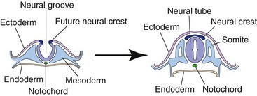
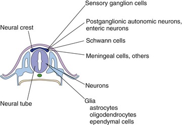
Cells of the neural crest grow at the apex of each neural fold. When the neural folds fuse to form the neural tube, the neural crest becomes a detached layer between the neural tube and the surface ectoderm (Fig. 2-1). Neural crest cells migrate from there, and go on to form most neurons and glial cells of the PNS (and much more). These include the sensory neurons of spinal and most cranial nerve ganglia, postganglionic autonomic neurons, and the Schwann cells of peripheral nerves and ganglia (Fig. 2-2). The neural tube goes on to form the CNS.
The Sulcus Limitans Separates Sensory and Motor Areas of the Spinal Cord and Brainstem
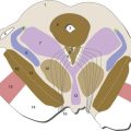
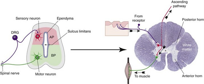
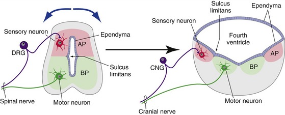
The sulcus limitans is a longitudinal groove that develops in the lateral wall of the embryonic spinal cord and extends into the rhombencephalon (the embryonic medulla and pons, as discussed a little later). It separates two groups of neuronal cell bodies, the alar plate (dorsal to the sulcus limitans in the spinal cord) and the basal plate (ventral to the sulcus limitans in the spinal cord). The alar and basal plates go on to become sensory and motor structures, respectively (Fig. 2-3). The spinal alar plate becomes the posterior horn, where primary sensory neurons terminate. The spinal basal plate becomes the anterior horn, where the cell bodies of motor neurons live.
The walls of the neural tube are spread apart in the rhombencephalon, forming the floor of the fourth ventricle, so in the medulla and pons the alar plate ends up lateral to the basal plate (Fig. 2-4). The same development into sensory and motor structures occurs, however, so cranial nerve sensory nuclei are lateral to cranial nerve motor nuclei in the adult brainstem (see Fig. 12-2).
The Neural Tube Has a Series of Bulges and Flexures
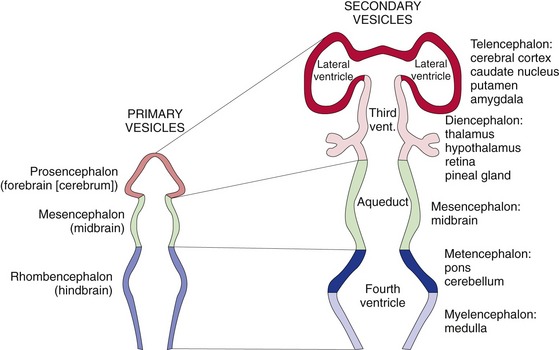
The prosencephalon and rhombencephalon each divides into two secondary vesicles, so there is a total of five secondary vesicles. The prosencephalon forms the telencephalon (“end-brain”) and the diencephalon (“in-between-brain”). The telencephalon gives rise to the two cerebral hemispheres, whose cavities become the lateral ventricles. The diencephalon gives rise to the thalamus, hypothalamus, retina, pineal gland, and several other structures; its cavity becomes the third ventricle. The mesencephalon remains undivided as the midbrain; its cavity persists as the cerebral aqueduct, which interconnects the third and fourth ventricles. The rhombencephalon forms the metencephalon and the myelencephalon, which together give rise to the cerebellum and the rest of the brainstem, and enclose the fourth ventricle. This ventricular arrangement is shown schematically in Fig. 2-5 and more realistically in THB6 Figures 5-1 and 5-2, pp. 100 and 101.
Bends in the neural tube also determine some aspects of the shape of the adult brain. Two bends are particularly important: the pontine flexure causes the walls of the neural tube to separate and form the floor of the fourth ventricle (see Fig. 2-4), and the cephalic flexure persists as the bend between the long axes of the prosencephalon (cerebrum) and the rest of the CNS (see Fig. 3-1).
Growth of the Telencephalon Overshadows Other Parts of the Nervous System
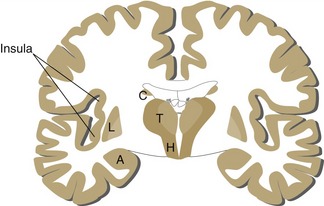
The telencephalon grows much more rapidly than the other vesicles. It folds down beside the diencephalon and fuses with it, and some cerebral cortex (the insula) develops over the site of fusion (Fig. 2-6). Subsequent growth of the cerebral hemisphere pivots about the insula, and the hemisphere grows around in a C-shaped arc through the parietal, occipital, and temporal lobes. This C shape is more than an anatomical curiosity that explains the shape of the lateral ventricles: in some cases, neural structures retain connections to distant sites in the forebrain, and C-shaped tracts interconnecting them are the result (THB6 Figure 3-18, p. 68).