CHAPTER 11 DERMATOPATHOLOGY
APPROACH TO PEDIATRIC SKIN BIOPSY INTERPRETATION


SPECIFIC ENTITIES IN PEDIATRIC DERMATOPATHOLOGY


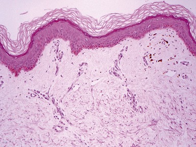
CUTIS LAXA
Genetics


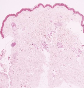
FOCAL DERMAL HYPOPLASIA (GOLTZ SYNDROME)


Clinical features


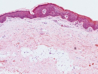


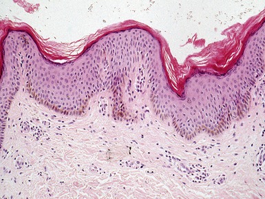
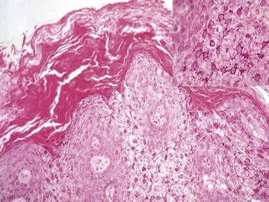
RESTRICTIVE DERMOPATHY


ICHTHYOSES
Introduction


ICHTHYOSIS VULGARIS


X-LINKED RECESSIVE ICHTHYOSIS


LAMELLAR ICHTHYOSIS / CONGENITAL ICHTHYOSIFORM ERYTHRODERMA (NON-BULLOUS ICHTHYOSIFORM ERYTHRODERMA)


Clinical features


BULLOUS ICHTHYOSIFORM ERYTHRODERMA / EPIDERMOLYTIC HYPERKERATOSIS


Genetics


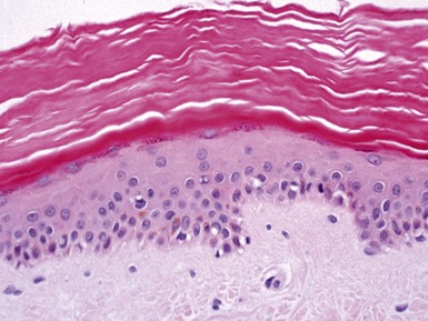
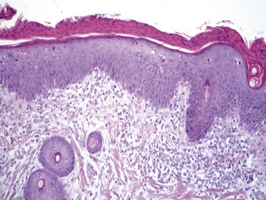
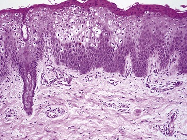
SJÖGREN–LARSSON SYNDROME


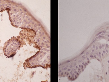
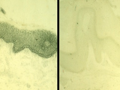
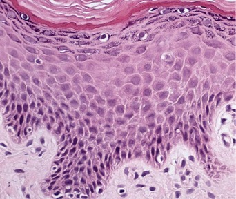
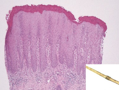
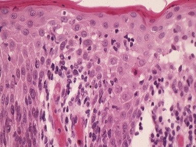
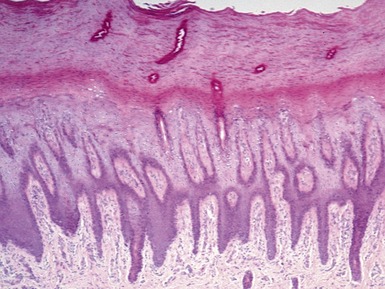
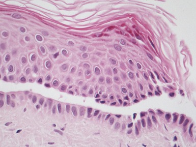
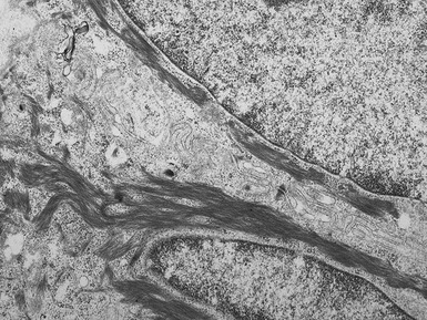
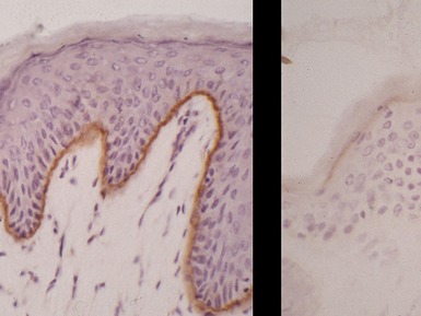
Histopathological features (Figs 11.14, 11.15)


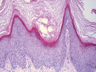
Fig 11.14 Photomicrograph of skin biopsy from a patient with Sjogren–Larsson syndrome demonstrating papillomatosis, acanthosis, hyperkeratosis and thickening of the granular layer.
NEUTRAL LIPID STORAGE DISEASE


NETHERTON’S SYNDROME


Clinical features


OMENN SYNDROME


KID SYNDROME (KERATITIS, ICHTHYOSIS AND DEAFNESS)


CONRADI–HUNERMANN–HAPPLE SYNDROME


CHILD SYNDROME (CONGENITAL HEMIDYSPLASIA WITH ICHTHYOSIFORM ERYTHRODERMA AND LIMB DEFECTS)


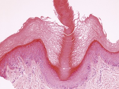
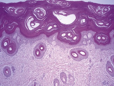
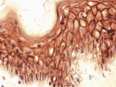
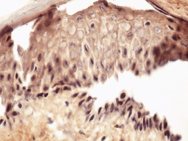
HEREDITARY PALMO-PLANTAR KERATODERMAS (PPKs)


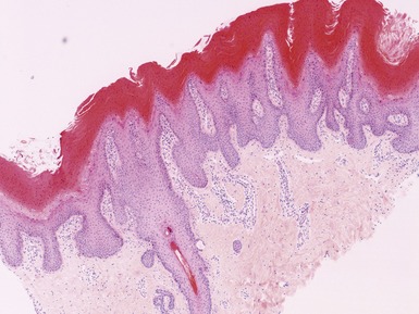
Histopathological features (Figs 11.23, 11.24)


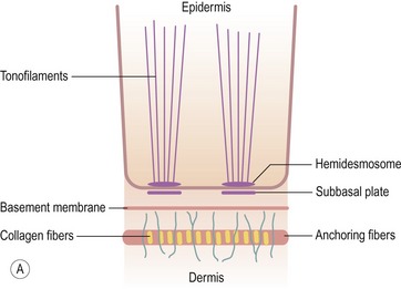
EPIDERMOLYSIS BULLOSA
Introduction


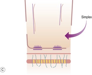
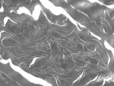
EPIDERMOLYSIS BULLOSA SIMPLEX


Clinical features


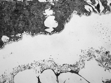
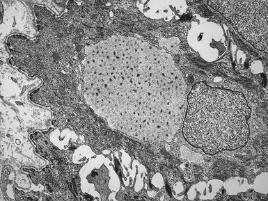
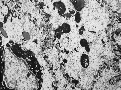
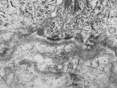
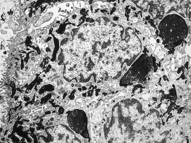
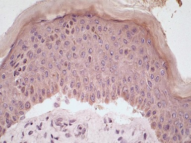
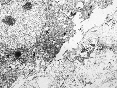
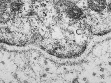
Histopathological features (Figs 11.27–11.35)


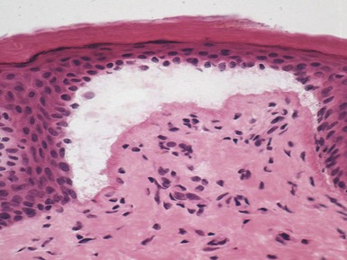
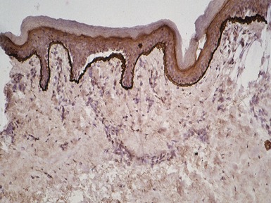
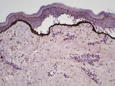
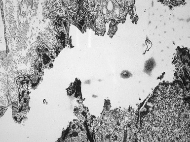
Fig 11.30 Electron micrograph of a skin biopsy from a patient with epidermolysis bullosa simplex demonstrating that the split is within the basal keratinocyte. A fragment of the keratinocyte can be seen in the lower part of the split.


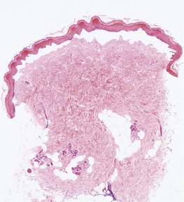
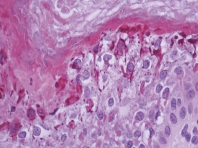
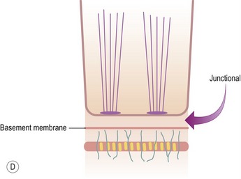
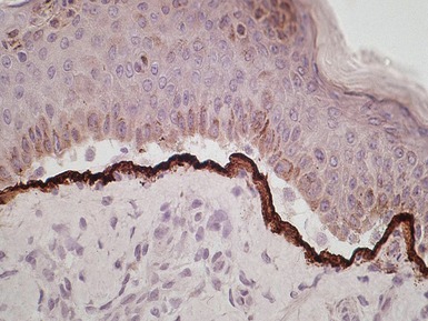
JUNCTIONAL EPIDERMOLYSIS BULLOSA


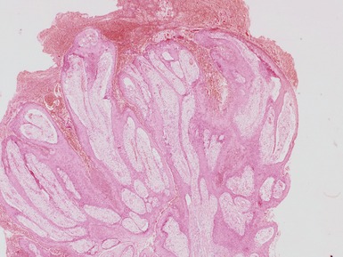
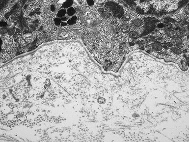
Histopathological features (Figs 11.36–11.43)


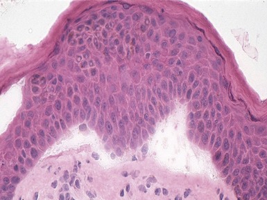
Fig 11.36 Photomicrograph of a frozen section of a skin biopsy from a patient with junctional epidermolysis bullosa demonstrating a split between the epidermis and the dermis.

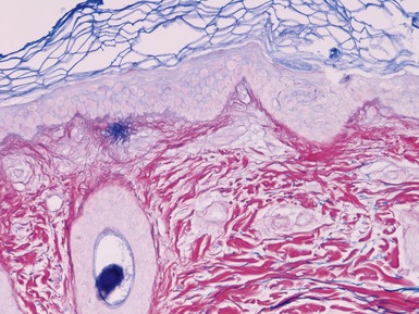
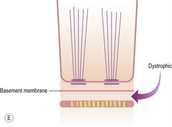
DYSTROPHIC EPIDERMOLYSIS BULLOSA


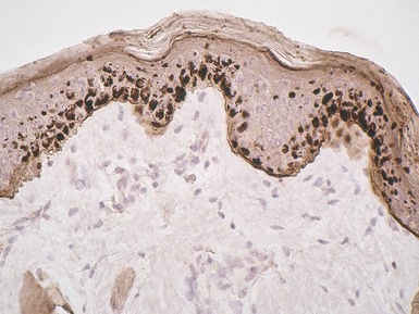
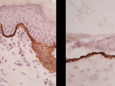
Histopathological features (Figs 11.44–11.49)


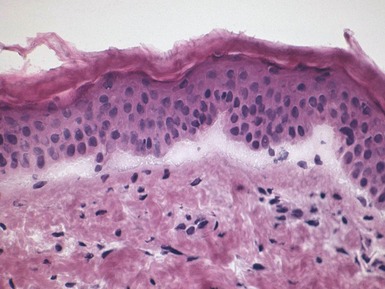
Fig 11.44 Photomicrograph of a frozen section of a skin biopsy from a patient with dystrophic epidermolysis bullosa demonstrating a split between the epidermis and the dermis.