63 Dermatomyositis
Salient features
History
• The adult form usually occurs after the age of 40 years
• Weakness of proximal muscles evolving over weeks or months: difficulty in getting from a low chair or squatting position, climbing stairs, lifting and running
• Dysphagia (caused by weakness of the muscles of the pharynx)
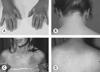
• Ask the patient whether the red rash is made worse by exposure to sunlight
Questions
What investigations would you like to do?
• Estimation of serum creatine kinase (also known as creatine phosphokinase) levels (mirrors disease activity)
• Electromyography shows myopathic changes (spontaneous fibrillation, salvos of repetitive potentials, and short duration of polyphasic potentials of low amplitude)
• Muscle biopsy (will show necrosis and phagocytosis of muscle fibres, and interstitial and perivascular infiltration of inflammatory cells)
• MRI and MR spectroscopy of affected tissue may show abnormal signals in striated muscle and abnormalities of muscle metabolism.
How would you treat such patients?
• Most patients respond to steroids; prednisolone is the first-line drug for the empirical treatment of polymyositis and dermatomyositis.
• Resistant dermatomyositis may benefit from methotrexate, azathioprine and high-dose intravenous immunoglobulin.
• Those with an underlying neoplasm may see the dermatomyositis remit after treatment of the tumour.
Advanced-level questions
How would you classify polymyositis–dermatomyositis?
The classification of Bohan et al. (Medicine 1977;56:255) is into five groups:
I: primary idiopathic polymyositis
II: primary idiopathic dermatomyositis
III: dermatomyositis (or polymyositis) associated with neoplasia (Mayo Clin Proc 1986;61:645)
IV: childhood dermatomyositis (or polymyositis) associated with vasculitis
V: polymyositis (or dermatomyositis) with associated collagen vascular disease.


