105
Dermatomyositis
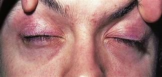
Violaceous hue and telangiectasias along the lid margin characterize the heliotrope of dermatomyositis.
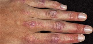
Flat-topped violaceous papules, Gottron’s papules, are seen on the knuckles and proximal phalangeal joints.
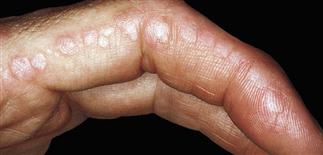
Gottron’s papules are flat-topped papules that may occur along the lateral fingers. They are pathognomonic for dermatomyositis.
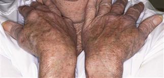
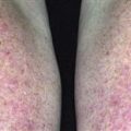
In this patient with dermatomyositis, a faint violaceous blush is seen along the extensor arms. The knuckles also have a characteristic violaceous hue.
DESCRIPTION
Acquired, idiopathic connective tissue disease characterized by proximal muscle weakness and characteristic violaceous skin rash prominent on eyelids, scalp, metacarpophalangeal joints, and bony prominences. Roughly 10% have skin findings without evidence of muscle disease. Amyopathic dermatomyositis is skin disease in absence of muscle disease over 6 months; 2 years required to confirm this.
HISTORY
• Twice as common in females. • At least 15% of cases occur in children. • May present with gradual onset of typical skin findings, or with a proximal muscle weakness. • Patients often have difficulty combing hair, ascending stairs, or rising from a chair without using arms. • Cutaneous lesions often precede muscle involvement by 3–6 months.
PHYSICAL FINDINGS
• Heliotrope rash refers to violaceous erythema of eyelids. • Pathognomonic Gottron’s papules located over bony prominences, particularly proximal interphalangeal, distal interphalangeal, and metacarpophalangeal joints. Violaceous papules and plaques. • Shawl sign refers to violaceous erythema over back of neck, posterior shoulders. • Poikiloderma (erythema, telangiectasia, hypopigmentation and hyperpigmentation, and atrophy) is typical finding in skin of upper chest, posterior shoulders, buttocks, and back. • Periungual changes: ragged cuticles, erythema, telangiectasia. • Scalp findings include alopecia, pruritus, erythema scaling. • Skin lesions often pruritic. • ‘Mechanic’s hands’ refers to scaly fissures and inflammatory changes on hands bilaterally. • Calcinosis rare but severe associated finding. • Muscle involvement: proximal and symmetric. • Dysphagia in 12–45%. Fatigue. • Elevation of muscle enzymes. Creatine kinase to follow disease activity. • Muscle biopsy or muscle imaging with magnetic resonance imaging. • May be a paraneoplastic disease; older patients at greater risk for associated malignancy. Screening and rescreening for malignancy is important in patients older than 50.
TREATMENT
• Broad-spectrum sunscreens, sun-protective clothing, behavior modification. • Earlier intervention, therapy with systemic corticosteroids results in improved prognosis if muscle disease. • Systemic corticosteroids (prednisone 0.5–1.5 mg/kg q.d.) are treatment cornerstone, although rash may not respond. • Hydroxychloroquine 200–400 mg q.d., alone or combined with quinacrine 100–200 mg q.d., results in skin improvement; response may be incomplete. • Steroid-sparing options for skin disease include immune suppressive agents methotrexate 2.5–30 mg/week, intravenous immunoglobulin 2 g/kg for 2 consecutive days. • Antihistamines for pruritus (non-sedating during day or sedating at night). • Second-line agents for muscle disease treatment and steroid-sparing: methotrexate 25–50 mg/week, azathioprine 2 mg/kg q.d., cyclosporine 5 mg/kg q.d., cyclophosphamide 2 mg/kg q.d., intravenous immunoglobulin 2 g/kg. • Passive range of motion exercise, rest, proper nutrition, physical and occupational therapy are important.