23. Dermatomyositis
Definition
Dermatomyositis is a disorder of collagen/connective tissue characterized by nonsuppurative skin inflammation and subcutaneous tissue and muscle fiber necrosis. It may be acute, subacute, or chronic.
Incidence
The most recent estimates indicate that dermatomyositis occurs at the rate of about 5.5:1,000,000. The frequency seems to be increasing. There is a 2:1 predilection toward females, but no apparent preference for a particular race.
Etiology
The cause of dermatomyositis is not fully understood. There are suggestions that it is caused by a complement-mediated vascular inflammation. The immunologic abnormalities may be triggered by a wide variety of agents.
Potential Triggers of Immunologic Abnormalities Related to Dermatomyositis
• Borrelia species
• Collagen injections
• Drug ingestion
• Silicone breast implants
• Toxoplasma species
• Viruses
Signs and Symptoms
• Arthralgia
• Arthritis
• Calcinosis of skin or muscle
• Dysphagia
• Dysphonia
• Dyspnea
• Dysrhythmias
• Facial erythema
• Gottron papules
• Heliotrope rash
• Joint swelling
• Malar erythema
• Muscle tenderness
• Muscle weakness
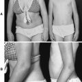
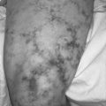
• Poikiloderma
• Pruritic skin lesions
• Psoriasis from dermatitis
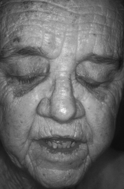 |
| Dermatomyositis. Note the rash affecting the eyelids. |
Medical Management
The muscle inflammation discomfort benefits from inactivity, such as bed rest. Physical therapy, on the other hand, helps counteract the muscle weakness and helps prevent contractures.
Some patients develop dysphagia and may be more prone to aspiration and aspiration pneumonia; the occurrence of both can be reduced by avoiding eating for several hours before bedtime. The incidence of reflux/aspiration can also be decreased by elevating the head of the bed.
Aside from bed rest and physical therapy, systemic corticosteroids are a standard of treatment. Prednisone is the typical first-line drug used. On establishing remission, the drug is tapered to discontinuation over time to reduce the probability of a relapse.
Chronic administration of corticosteroids often results in the development of toxic effects. These effects are frequently counteracted by initiation of an immunosuppressive or cytotoxic drug. Methotrexate is usually the drug of choice for the steroid-sparing medication; however, azathioprine and mycophenolate mofetil are viable alternatives. Intravenous administration of high-dose immunoglobulin is reserved for the patient for whom steroids and/or immunosuppressant/cytotoxic therapy proves unsuccessful. It is carried out monthly, and generally produces only short-term benefits.
The cutaneous aspects of this disease are the most difficult to treat. The first-line intervention is for the patient to realize and understand the photosensitive nature of the disease. The patient should limit exposure to the sun as much as possible, either by outright avoidance or by application of copious amounts of sunscreen solution. Other practical actions include wearing broad-brimmed hats, using umbrellas, and covering skin with clothing. There have been reports of beneficial effects of hydroxychloroquine and chloroquine on the cutaneous manifestations of this disease.
Calcinosis is debilitating if it becomes established. Remission is possible, but if it occurs, it is only after the patient has had the disease for years. This complication has reportly been gradually resolved by administration of the calcium channel–blocking drug, diltiazem.
Complications
• Aspiration
• Aspiration pneumonia
• Calcinosis
• Cardiac conduction abnormalities
• Congestive heart failure
• Contractures
• Dysphagia
• Gastrointestinal ulcers and perforations
• Interstitial lung disease
• Myocarditis
• Necrotizing vasculitis
Anesthesia Implications
Dysphagia increases the potential for aspiration, particularly during induction of general anesthesia. Prudence dictates that the patient receive a rapid sequence induction.
Muscle weakness is a hallmark of this disease. It is especially evident in the neck flexors and shoulder and pelvic girdles. Administration of muscle relaxants may need to be reduced because these muscle groups exhibit greater sensitivity to them. On completion of anesthesia, even though reversal is deemed complete, the patient may not be able to produce a sustained head-lift because of the already weak neck muscles.
Interstitial lung disease may proceed to restrictive lung disease. As a result, diffusing capacity is diminished and the potiential for air trapping increases, as does the potential for barotrauma. When positive pressure ventilation is required, peak inspiratory pressures should be reduced while expiratory time is increased to reduce the potential for lung injury.
The patient’s respiratory capabilities should be thoroughly assessed preoperatively utilizing pulmonary function testing, in particular vital capacity, arterial blood gas analysis, and chest x-rays. If the patient’s capabilities are deemed marginal, the patient should not be allowed to spontaneously breathe either intra- or postoperatively. In such cases general anesthesia would be the more appropriate choice for anesthesia technique. Regional anesthesia should be used in combination with general anesthesia. Postoperative mechanical ventilatory should be anticipated for the patient with dermatomyositis who receives general anesthesia.
Cardiac conduction abnormalities increase the potential for development of detrimental dysrhythmias.
Chronic administration of corticosteroids requires a perioperative dose of steroid before surgical incision.
Renal function should be assessed preoperatively. The patient with dermatomyositis may develop necrotizing vasculitis, which may affect renal function.