[level-membership-for-dermatology-category]
Chapter 64 Dermatologic emergencies
Vesiculobullous disorders and drug reactions
Table 64-1. Clinicopathologic Features of Toxic Epidermal Necrolysis (TEN) versus Stevens-Johnson Syndrome (SJS)
| TEN | SJS | |
|---|---|---|
| Maximal intensity | 1–3 days | 7–15 days |
| Skin pain | Severe | Minimal |
| Mucosal involvement | Mild | Severe |
| Lesional pattern | Diffuse erythema, desquamation | Annular and targetoid lesions |
| Skin histology | Few inflammatory cells | Numerous inflammatory cells |
| Prognosis | Poor | Excellent |
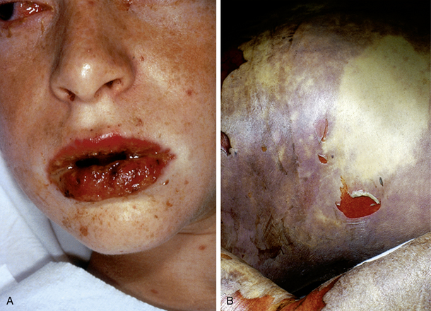
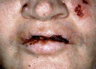
Figure 64-2. Pemphigus vulgaris demonstrating erosive lesions of the lips and left cheek.
(Courtesy of James E. Fitzpatrick, MD.)
Groves RW: Pemphigus: a brief review, Clin Med 9: 371–375, 2009.
Table 64-2. Diagnostic Signs in Dermatologic Infectious Emergencies
| Petechial/Palpable Purpura |
Key Points: Dermatologic Emergencies
Inflammatory cutaneous disorders
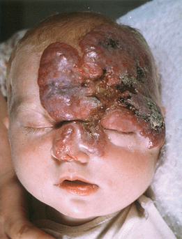
Most commonly, these tumors are only a cosmetic problem, but if they occur around the eyes or in the oral cavity (Fig. 64-6), they can cause significant morbidity and mortality. Some ophthalmologists suggest that even a few days of obstructed vision in a newborn can inhibit normal visual development. Therefore, an infantile hemangioma that may block an infant’s visual fields should be treated aggressively. Likewise, enlarging infantile hemangiomas of the upper respiratory tract and oral cavity can result in acute emergent situations and must be treated early in their course. In rare cases, large hemangiomas can also produce high-output cardiac failure.
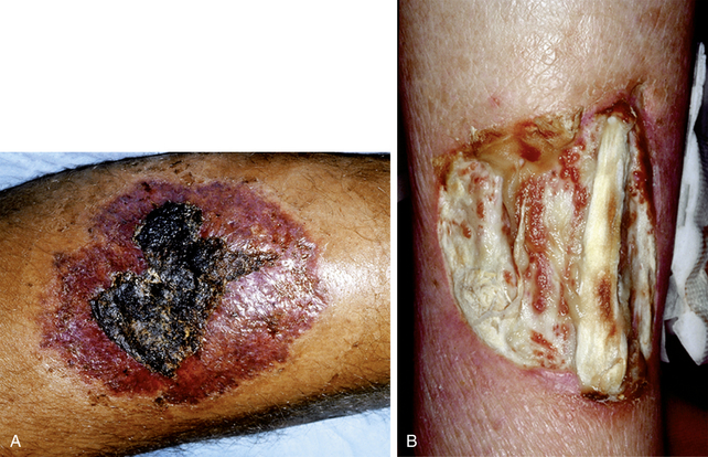
Manganoni AM, Venturini M, Scolari F, et al: The importance of skin biopsy in the diverse clinical manifestations of cholesterol embolism, Br J Dermatol 150:1230–1231, 2004.
[/level-membership-for-dermatology-category][not-level-membership-for-dermatology-category]
Chapter 64 Dermatologic emergencies
Vesiculobullous disorders and drug reactions
Table 64-1. Clinicopathologic Features of Toxic Epidermal Necrolysis (TEN) versus Stevens-Johnson Syndrome (SJS)
| TEN | SJS | |
|---|---|---|
| Maximal intensity | 1–3 days | 7–15 days |
| Skin pain | Severe | Minimal |
| Mucosal involvement | Mild | Severe |
| Lesional pattern | Diffuse erythema, desquamation | Annular and targetoid lesions |
| Skin histology | Few inflammatory cells | Numerous inflammatory cells |
| Prognosis | Poor | Excellent |
Figure 64-2. Pemphigus vulgaris demonstrating erosive lesions of the lips and left cheek.
(Courtesy of James E. Fitzpatrick, MD.)
Groves RW: Pemphigus: a brief review, Clin Med 9: 371–375, 2009.
Table 64-2. Diagnostic Signs in Dermatologic Infectious Emergencies
| Petechial/Palpable Purpura |
[/not-level-membership-for-dermatology-category]
