Derivatives of the skin
Hair
There are three types of hair:
1. Lanugo hairs are fine and long, and are formed in the fetus at 20 weeks’ gestation. They are normally shed before birth, but may be seen in premature babies.
2. Vellus hairs are the short, fine, light-coloured hairs that cover most body surfaces.
3. Terminal hairs are longer, thicker and darker, and are found on the scalp, eyebrows, eyelashes and also on the pubic, axillary and beard areas. They originate as vellus hair; differentiation is stimulated at puberty by androgens.
Structure
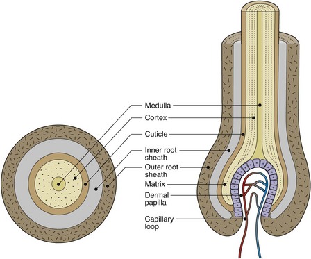
The hair follicle is an invagination of the epidermis containing a hair. The portion above the site of entry of the sebaceous duct is the infundibulum. The hair shaft consists of an outer cuticle that encloses a cortex of packed keratinocytes with (in terminal hairs) an inner medulla (Fig. 1). The germinative cells are in the hair bulb; associated with these cells are melanocytes, which synthesize pigment. The arrector pili muscle is vestigial in humans; it contracts with cold, fear and emotion to erect the hair, producing ‘goose pimples’.
Nails
Structure
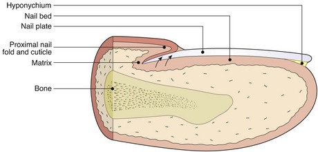
The nail matrix contains dividing cells which mature, keratinize and move forward to form the nail plate (Fig. 2). The nail plate has a thickness of 0.3–0.5 mm and grows at a rate of 0.1 mm/24 h for the fingernail. Toenails grow more slowly. The nail bed, which produces small amounts of keratin, is adherent to the nail plate. The adjacent dermal capillaries produce the pink colour of the nail; the white lunula is the visible distal part of the matrix. The hyponychium is the thickened epidermis that underlies the free margin of the nail.
Sebaceous glands
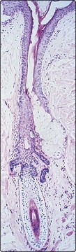
Sebaceous glands are found associated with hair follicles (Fig. 3), especially those of the scalp, face, chest and back, and are not found on non-hairy skin. They are formed from epidermis-derived cells and produce an oily sebum, the function of which is uncertain. The glands are small in the child, but become large and active at puberty, being sensitive to androgens. Sebum is produced by holocrine secretion in which the cells disintegrate to release their lipid cytoplasm.
Sweat glands
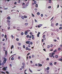
Sweat glands (Fig. 4) are tube-like and coiled glands, located within the dermis, which produce a watery secretion. There are two separate types: eccrine and apocrine.
Other structures in skin
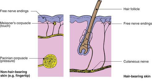
Nerve supply
The skin is richly innervated (Fig. 5), with the highest density of nerves being found in areas such as the hands, face and genitalia. All nerves supplying the skin have their cell bodies in the dorsal root ganglia. Both myelinated and non-myelinated fibres are found. The nerves contain neuropeptides, e.g. substance P.
Blood and lymphatic vessels
The skin also has a rich and adaptive blood supply. Arteries in the subcutis branch upwards, forming a superficial plexus at the papillary/reticular dermal boundary. Branches extend to the dermal papillae (Fig. 6), each of which has a single loop of capillary vessels, one arterial and one venous. Veins drain from the venous side of this loop to form the mid-dermal and subcutaneous venous networks. In the reticular and papillary dermis, there are arteriovenous anastomoses that are well innervated and concerned with thermoregulation (see p. 7).
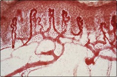
Fig. 6 Superficial dermal blood vessels.
Capillary loops branch off the superficial vascular plexus and extend into each dermal papilla.


