Hand Deformities
Impairment of hand function can lead to severe disability.
Causes
Congenital
Acquired
• Rheumatoid arthritis (Fig. 32)
• Trauma (mal-united fracture)
• Burns
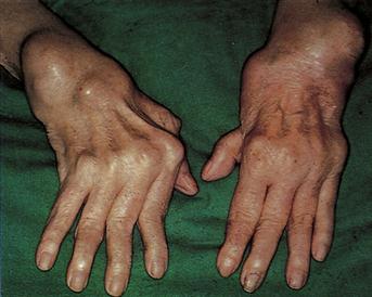
Figure 32 Rheumatoid hand.
Early changes include synovitis of the wrist, metacarpophalangeal joints and proximal interphalangeal joints. In severe advanced changes, there is massive tendon swelling over the dorsal surfaces of the wrists, severe muscle wasting, ulnar deviation of the metacarpophalangeal joints and swan-neck deformity of the fingers.
History
Congenital
These lesions will be recognisable at birth. Arthrogryposis multiplex congenita is due to non-differentiation of mesenchymal tissues, often together with a neurogenic element, the combination producing gross contractures.
Acquired
Trauma and burns
There will be a clear history of previous trauma and burns.
Dupuytren’s contracture
In the early phases, there will be little deformity, the patient merely complaining of thickening in the palm near the base of the ring finger. Eventually, the patient will complain of being unable to straighten the ring and little fingers. The grip is also affected. Check for a history of epilepsy, cirrhosis or diabetes.
Rheumatoid arthritis
By the time the hand is deformed, the patient is usually well aware of the diagnosis. There is pain and swelling of the joints with weakness of the hand, together with gross deformity.
Volkmann’s ischaemic contracture
This is shortening of the long flexor muscles of the forearm due to ischaemia. There may be a history of trauma, e.g. supracondylar fracture of the humerus, a tight plaster restricting blood flow, or arterial disease, e.g. embolism. Movements of the fingers are painful and limited. There may be ‘pins and needles’ in the hand due to pressure on nerves. Eventually a claw hand results.
Spinal cord lesions
By the time deformity of the limb has resulted, the diagnosis will usually be apparent. There may be a history of poliomyelitis.
Brachial plexus lesions
There will usually be a history of trauma, although occasionally, invasion of the brachial plexus from tumours may occur.
Peripheral nerve lesions
There is usually a clear history of trauma to suggest a nerve lesion. Check for any possible causes of peripheral neuropathy, e.g. diabetes, uraemia, connective tissue disease, leprosy.
Ollier’s disease
This results in deformities of the limb. There are irregularly distributed areas of cartilage and bone. The patient will complain of deformity of joints and bones and possible shortening of the limb, and will have noticed nodules along the line of bone.
Examination
Congenital
Congenital anomalies will be obvious at birth. They may be associated with congenital anomalies elsewhere.
Acquired
Trauma and burns
A variety of deformities may be seen, depending upon the degree and type of trauma. With burns, scarring of the skin will be obvious.
Dupuytren’s contracture
Examination will reveal a firm nodule in the palmar fascia near the base of the ring finger. Puckering of the skin is usually present around the nodule. The MCP joint and the PIP joint are flexed, the DIP joint being extended. The ulnar digits (ring and little finger) are most commonly affected.
Rheumatoid arthritis
There is thickening of the joints, especially the MCP joint and PIP joints. The joints become fusiform in shape. There is ulnar deviation of the fingers. The wrist joint develops a fixed flexion deformity with some ulnar deviation. Swan-neck and boutonnière deformities of the fingers will be apparent. Ultimately, tendon rupture occurs and this leads to a variety of deformities.
Volkmann’s ischaemic contracture
The skin of the hand is pale and the hand clawed and wasted. Extension of the fingers is limited but improves if the wrist is flexed. Eventually, the forearm muscles feel hard and taut due to fibrosis.
Spinal cord lesions
In the case of poliomyelitis, there is usually a clear history and the limb usually looks reddish-blue, wasted and deformed. Syringomyelia may cause a variety of hand deformities. With upper motor neurone lesions, there may be fixed flexion of the wrist and fingers with adduction of the thumb.
Brachial plexus injuries
Upward traction on the arm may damage the lowest root (T1) of the brachial plexus, which is the segmental supply of the intrinsic muscles of the hand. The hand becomes clawed (Klumpke’s paralysis). Check for a possible associated Horner’s syndrome due to traction of the cervical sympathetic chain.
Peripheral nerve lesions
With ulnar nerve lesions, it is appropriate to check for damage to the medial epicondyle. Also check for any evidence of recent lacerations at the wrist. With division at the wrist, all the intrinsic muscles of the fingers (except for the radial two lumbricals – median nerve) are paralysed and the hand appears clawed. The clawing is less for the index and middle fingers because the lumbricals are intact. In late cases, wasting of the interossei is clearly seen on the dorsum of the hand. There is sensory loss over the medial 1{1/2} fingers. If the nerve is injured at the elbow, the flexor digitorum profundus to the ring and little fingers is paralysed so that the clawing of these two digits is not so pronounced.
In median nerve lesions at the wrist, the thenar eminence becomes wasted due to paralysis of opponens pollicis and sensation is lost over the lateral 3{1/2} digits. Fine movements, e.g. picking up a small object, are clumsy. With lesions at the elbow, wrist flexion is weak and the forearm muscles wasted. Ulnar deviation occurs at the wrist, as the wrist flexion depends upon flexor carpi ulnaris and the medial half of flexor digitorum profundus. Often the hand is held with the medial two fingers flexed and the lateral two fingers straight.
Ollier’s disease
Multiple lumps (chondromas) are located on the bones. Often associated with limb shortening. Only one limb or one bone may be involved.
General Investigations
Most of the conditions described above will be obvious on clinical examination.
■ FBC, ESR
Hb ↓ in anaemia of chronic disease, e.g. rheumatoid arthritis.
■ CRP
↑ in inflammatory disease
■ Rheumatoid factor
Rheumatoid arthritis.
■ LFTs
Cirrhosis (Dupuytren’s contracture).
■ Blood glucose
Diabetes. Peripheral neuropathy, Dupuytren’s contracture.
■ Arm X-ray
Local fractures, e.g. evidence of previous supracondylar fracture of the humerus with subsequent Volkmann’s ischaemic contracture.
■ Hand X-ray
Rheumatoid arthritis. Ollier’s disease – multiple translucent areas in the bones of the hand. Evidence of mal-united fracture following trauma.
■ MRI
Spinal lesions, e.g. syringomyelia.
■ Nerve conduction studies
Brachial plexus and peripheral nerve lesions.