D
D-15 test See test, Farnsworth.
dacryoadenitis Inflammation of the lacrimal gland. The acute type is characterized by localized pain, swelling and redness over the upper temporal area of the eye. The chronic type is painless and develops slowly. A frequent cause is an associated systemic infection such as mumps, infectious mononucleosis, influenza, or it can be due to a local condition such as trachoma, herpes zoster or staphylococcal infection. The chronic type may be due to any of the granulomatous diseases (tuberculosis, syphilis, sarcoidosis). Treatment consists mainly of warm compresses and antibiotics.
See syndrome, Mikulicz’s.
dacryocele, congenital A congenital condition in which the infant is born with a swollen lacrimal sac filled with mucoid material. Physical examination reveals a bluish mass located in the nasal canthal region, probably due to an obstruction of the lower end of the nasolacrimal duct, with associated blockage of the canaliculi and puncta. Treatment includes antibiotics as well as nasolacrimal probing and irrigation in many cases. Syn. dacryocystocele.
dacryocystectomy Surgical removal of the lacrimal sac.
dacryocystitis Inflammation of the lacrimal sac. It is a rare condition, which may occur when there is a blockage of the nasolacrimal drainage system. The acute type gives rise to redness, tenderness and swelling below the lid margin, while in the chronic type there is epiphora and with pressure on the lacrimal sac pus will come out of the punctum. Treatment includes broad-spectrum antibiotics and warm compresses but surgery may be needed in the chronic type.
See fistula, lacrimal; lacrimal apparatus.
dacryoliths Concretions found in the lacrimal apparatus, in the puncta or canaliculi which it may occlude. The concretions are usually composed of epithelial cells, lipid, nonspecific debris as well as calcium.
dacryoma 1. A tumour or swelling anywhere within the lacrimal apparatus. 2. A blockage of a lacrimal punctum.
dacryops 1. A chronic watery eye. 2. A cyst in a tear duct of the lacrimal gland.
See epiphora.
Dalen–Fuchs nodules See Dalen– Fuchs nodules.
Dalrymple’s sign See sign, Dalrymple’s.
daltonism Term used formerly to designate colour blindness, usually deutan, so named because John Dalton (1766–1844) was the first to describe his own anomaly.
dapiprazole See alpha-adrenergic antagonist.
dark adaptation See adaptation, dark.
dark current See electrooculogram.
dark filter test See test, neutral density filter.
dark focus See accommodation, resting state of.
dark room test See test, provocative.
dark vergence See vergence, tonic.
dark wedge test See test, Bielchowsky’s phenomenon.
day blindness See hemeralopia.
daylight, artificial Illumination produced by a source of artificial light having a spectral distribution similar to that of daylight. CIE Illuminant C is considered to almost fulfill this criterion.
daylight, natural Illumination dependent on the sun and the extent of clear sky.
daylight vision See vision, photopic.
deaf-blind A person who has a severe hearing impairment in addition to a visual defect. It is usually congenital but it may result from ageing or some systemic disease or as part of a syndrome (e.g. Usher’s syndrome which accounts for about half of all cases of deaf-blind people; rubella syndrome).
debility The state of being feeble or without strength.
debridement Removal of dead or infected tissue or foreign material until surrounding healthy tissue is exposed. This is done to facilitate healing. Corneal debridement is usually performed with a cotton-tipped applicator, a spatula or with a sharp instrument. Example: debridement of some of the corneal epithelium in dendritic keratitis or in corneal erosion.
decentration (dec) A displacement, horizontal and/or vertical, of the centration point of a spectacle lens from the standard optical centre position (British Standard).
See centre, standard optical position; point, centration.
decibel (dB) 1. Unit used for the measurement of the intensity of a sound. 2. Light intensities are often presented on a logarithmic (rather than linear) scale. This is done, in particular, to abbreviate large numbers. Moreover, it has become common, especially in perimetry, to use decibels rather than log units. A decibel scale is a logarithmic scale where 10 decibels are equal to 1 log unit; 20 decibels, to 2 log units, etc. In perimetry, decibels are used to indicate the attenuation of brightness of the stimulus. Thus, a 20 dB stimulus is equal to one-tenth the brightness of a 10 dB stimulus.
decompensation Failure of an organ to fulfill its function adequately. Examples: corneal decompensation following years of extended contact lens wear; a failure of the eye movement system to overcome a heterophoria.
decongestant, ocular A pharmaceutical agent used to reduce hyperaemia in the eye, usually by vasoconstriction. Decongestants are weak concentrations of sympathomimetic or alpha-adrenergic agonists. Examples: adrenaline (epinephrine); naphazoline hydrochloride; phenylephrine hydrochloride, tetrahydrozoline hydrochloride. A decongestant can also be used to differentiate between conjunctival and ciliary injection; if the instillation of a decongestant alleviates eye redness the injection is primarily conjunctival, otherwise the redness is of ciliary origin.
decussation Crossing of nerve fibres passing through the mid-sagittal plane of the central nervous system and connecting with structures on the opposite side. Partial decussation occurs at the optic chiasma.
defocus To put or go out of focus.
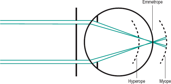
hyperopic d. State of the eye in which the retinal image is focused behind the retina. It may occur when placing a negative lens in front of an emmetropic presbyopic eye; in an uncorrected presbyopic hyperope; in a high hyperope unable to overcome the ametropia by accommodating; or as an accommodative lag in an uncorrected or corrected myope, as well as in an emmetrope viewing a near object.
See accommodation, lag of; over-correction.
myopic d. State of the eye in which the retinal image is focused in front of the retina. It may occur when placing a positive lens in front of an emmetropic eye; in an uncorrected or undercorrected myopic eye; or if there is a lead of accommodation, as is the case in some individuals viewing a distant object.
See accommodation, lead of; undercorrection.
degeneration Deterioration of tissue or organ resulting in reduced efficiency. Examples: degeneration of the cornea; degeneration of the retina.
age-related macular d. See macular degeneration, age-related.
cobblestone d. See degeneration, paving-stone.
Doyne’s honeycombed d. See drusen, familial dominant.
lattice d. of the retina See retina, lattice degeneration of the.
lipid droplet d. See keratopathy, actinic.
paving-stone d. Discrete, yellowish round areas of retinal thinning and depigmentation located near the ora serrata. The underlying choroid may be seen. It is a benign degeneration occurring with advancing age. Syn. cobblestone degeneration; peripheral chorioretinal degeneration.
pellucid marginal d. A rare condition characterized by bilateral, slowly progressive thinning and protrusion of the inferior peripheral cornea. The involved area is clear (hence the word pellucid), but the condition may be complicated by hydrops and the central cornea typically develops against the rule astigmatism. Treatment usually consists of gas permeable scleral lenses, but keratoplasty may be necessary.
See ectasia, corneal; hydrops; keratoconus.
peripheral chorioretinal d. See degeneration, paving-stone.
peripheral cystoid d. A degenerative process in the peripheral retina that occurs almost universally in the elderly. It consists of numerous, discrete cystic spaces in the outer plexiform or inner nuclear layer presenting a frothy appearance. The degeneration starts at the ora serrata and slowly progresses to the peripheral retina. If the cysts should join together, degenerative retinoschisis develops. It is not usually associated with retinal tears. The condition does not require any treatment.
Salzmann’s nodular d. A degenerative condition characterized by bluish-white, elevated nodules on the surface of the cornea. It may occur in people previously affected by trachoma, phlyctenular keratitis, vernal keratitis or keratoconjunctivitis sicca. Most cases are asymptomatic, but if the nodules impair vision, keratoplasty may be necessary.
senile macular d. See macular degeneration, age-related.
tapetoretinal d. A hereditary degeneration affecting the photoreceptors of the retina or the pigment epithelium layer. Some authors also include the choroid. Syn. tapetoretinopathy.
See choroideremia; retinitis pigmentosa.
Terrien’s marginal d. See ectasia, corneal.
vitreoretinal d. See disease, Wagner’s; syndrome, Stickler’s.
dellen A transient shallow depression in the cornea near the limbus which is caused by a local dehydration of the corneal stroma, leading to a compression of its lamellae. It can occur as a result of strabismus surgery, cataract surgery, swelling of the limbus (as in episcleritis or pterygium), rigid contact lens wear or senility.
DEM test See test, developmental eye movement.
demecarium bromide See anticholinesterase.
dendritic keratitis See keratitis, dendritic.
densitometry, retinal A technique used to study visual pigments in vivo. It consists of measuring the small fraction of light that is reflected by the pigment epithelium of the retina before and after bleaching with a bright source of light.
density An indication of the compactness of a substance. It is expressed as the ratio of the mass of the substance to its unit volume. The common units are g/cm3 and kg/m3. This property is usually given by lens manufacturers, the greater the density of a material, the greater its weight, all other factors being equal.
density, optical A term applied to optical filters. It is equal to the logarithm to the base 10 of the reciprocal of the transmission factor T thus,
< ?xml:namespace prefix = "mml" />
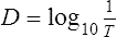
where D is the symbol for optical density. Syn. absorbance.
See absorbance; absorption; spectrophotometer; transmittance.
Table D1
Density of optical lens materials
| n | density (g/cm3) | |
| glass | ||
| spectacle crown | 1.523 | 2.54 |
| dense barium crown | 1.620 | 3.71 |
| dense flint | 1.706 | 3.20 |
| dense barium flint | 1.700 | 4.10 |
| titanium oxide | 1.701 | 2.99 |
| plastics | ||
| PMMA | 1.490 | 1.19 |
| CR-39 | 1.498 | 1.32 |
| polycarbonate | 1.586 | 1.2 |
| cellulose acetate | 1.48 | 1.2 |
| butyrate | ||
| cellulose propionate | 1.46 | 1.2 |
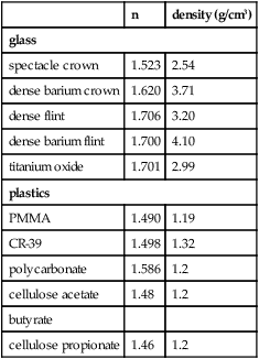
Table D2
Relationship between optical density D and light transmission T of optical filters
| D | T (%) |
| 0.0 | 100 |
| 0.1 | 79.4 |
| 0.2 | 63.1 |
| 0.3 | 50.1 |
| 0.4 | 39.8 |
| 0.5 | 31.6 |
| 0.6 | 25.1 |
| 0.7 | 20.0 |
| 0.8 | 15.8 |
| 0.9 | 12.6 |
| 1.0 | 10.0 |
| 1.5 | 3.16 |
| 2.0 | 1.0 |
| 2.5 | 0.32 |
| 3.0 | 0.1 |
| 4.0 | 0.01 |
Denver Developmental Screening Test See test, developmental and perceptual screening.
deorsumduction See depression.
deorsumvergence See infravergence.
deorsumversion See version.
depolarization A change in the value of the resting membrane potential towards zero. The inside of the cell becomes less negative compared to the outside. This is due to a change in permeability and migration of sodium ions into the interior of the cell. Depolarization is excitatory because the membrane potential shifts towards the neuron’s threshold at which an action potential occurs.
See hyperpolarization; synapse.
depolished glass See glass, ground.
deposits, contact lens Accumulation of materials on or into the matrix of contact lenses. They are mainly tear components (proteins, lipids, mucin, calcium) but other materials can be found (e.g. mercurial or iron deposits, nicotine, hand cream). Deposits reduce comfort, vision, patient tolerance and discolour and spoil the lenses. They may act as antigens for the development of giant papillary conjunctivitis. Most of these deposits can be removed with a surfactant, an enzymatic system and a calcium-preventing solution.
depression Downward rotation of an eye. It is accomplished by the inferior rectus and superior oblique muscles. It can be induced by using base-up prisms. Syn. infraduction; deorsumduction.
depressors Extraocular muscles that move the eye downward, such as the inferior rectus and the superior oblique.
deprivation amblyopia See amblyopia, deprivation.
deprivation, sensory The condition produced by a loss of all or most of the stimulation from the visual, auditory, tactile and other sensory systems. Often, deprivation involves only one modality (e.g. vision). Methods used for deprivation include diffusing goggles, white noise, padded gloves, etc. Its effect has shown the necessity of continuous sensory activity to maintain the normal development and functioning of any sensory system.
deprivation, visual The condition produced by a loss of form vision. It may occur as a result of an anomaly within the eye (e.g. opacification of the cornea), or it can be artificially induced (e.g. by placing a transparent plastic occluder in front of the eye, as used in myopia research with animals).
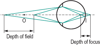
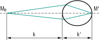
depth of field For a given setting of an optical system (or a steady state of accommodation of the eye) it is the distance over which an object may be moved without causing a sharpness reduction beyond a certain tolerable amount. Depth of field increases when the diaphragm (or pupil) diameter diminishes as, for example, in old eyes (Fig. D1). Examples: viewing at infinity, the depth of field ranges between infinity and about 3.6 m for a pupil of 4 mm in diameter; and between infinity and about 2.3 m for a 2 mm pupil. At a viewing distance of 1 m, the depth of field ranges from about 1.4 m to 80 cm with a 4 mm pupil; and from about 1.8 m to 70 cm with a 2 mm pupil.
See distance, hyperfocal.
depth of focus For a given setting of an optical system (or a steady state of accommodation of the eye) it is the distance in front and behind the focal point (or retina) over which the image may be focused without causing a sharpness reduction beyond a certain tolerable amount. (The criterion could be as much as a line of letters on a Snellen chart.) The depth of focus D is represented by the total distance in front and behind (Fig. D1). As with depth of field, it is inversely proportional to the diameter of the diaphragm (or pupil). It can be calculated, expressed in dioptres, using the equation
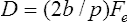
where Fe is the power of the emmetropic eye, p the pupillary diameter and b the maximum size of the retinal image beyond which it is perceived as blurred. Example: Assuming a pupil of 3 mm and a retinal image spread over five cones, each 0.002 mm in diameter and spaces between the cones of 0.0005 mm, that is a total size = 0.012 mm, D = 2 × 0.012/3 × 60 = 0.48 D. Thus an object appears clear if the vergence at the eye varies in the range ±0.24 D. If the eye is focused at infinity, the equation becomes D = (b/p) Fe.
depth perception See perception, depth.
depth, vertex See vertex depth.
dermatitis See eczema.
dermatochalasis A condition in which there is a redundancy of the skin of the upper eyelids. It is often associated with a protrusion of fat through a defective orbital septum. The condition occurs usually in old people. The excess skin may cause pseudoptosis. In severe cases it may obstruct vision. Treatment is surgical. Syn. ptosis adiposa; ptosis atrophica.
See blepharochalasis.
dermoid cyst See cyst, dermoid.
desaturated D–15 test See test, Farnsworth.
Descartes’ law See law of refraction.
Descemet’s membrane See membrane, Descemet’s.
descemetocele A forward bulging of Descemet’s membrane due to either trauma or a deep corneal ulcer which has eroded the overlying stroma. Syn. keratocele.
See keratolysis.
desiccation The process of becoming dry. See eye, dry.
desmosome A site of adhesion between two adjacent cells, such as in the corneal epithelium. It consists of a small, dense body in which the two halves are separated by an intercellular gap filled with extracellular substance. The basal cells are attached at irregular intervals to the underlying basement membrane (adjacent to Bowman’s layer) by hemidesmosomes (one half of a desmosome). Thus, scraping off the epithelium usually leaves fragments of the basal cells attached to the basement membrane.
detachment, choroidal See choroidal detachment.
detachment, retinal See retinal detachment.
deturgescence State of relative dehydration maintained by the normal cornea that is necessary for transparency. It is maintained by the epithelium, which to a large extent is impermeable to water, and also by a metabolic transport system in the endothelium.
deutan A person who has either deuteranomaly or deuteranopia.
deuteranomal Person who has deuteranomaly.
deuteranomaly A type of anomalous trichromatism in which an abnormally high proportion of green is needed when mixing red and green light to match a given yellow. This is the most common type of colour vision deficiency, occurring in about 4.6% of males and 0.35% of the female population. Syn. deuteranomalous trichromatism; deuteranomalous vision; green-weakness.
See anomaloscope; colour vision, defective.
deuteranope Person who has deuteranopia.
deuteranopia Type of dichromatism in which red and green are confused, although their relative spectral luminosities are practically the same as in normals. In the spectrum, the deuteranope only sees two primary colours, the long wavelength portion of the spectrum (yellow, orange or red) appears yellowish and the short wavelength portion (blue or violet) appears bluish. There is, in between, a neutral point which appears whitish or colourless, at about 498 nm. It occurs in slightly over 1% of the male population and only rarely in females. Syn. green blindness (although this term is incorrect as green lights appear to a deuteranope as bright as to a normal observer).
See colour vision, defective; dichromatism.
developmental and perceptual screening test See test, developmental and perceptual screening.
deviating eye See eye, deviating.
deviation 1. In strabismus, the departure of the visual axis of one eye from the point of fixation. 2. A change in direction of a light ray resulting from reflection or refraction at an optical surface.
angle of d. See angle of deviation.
conjugate d. The simultaneous and equal rotations of the eyes in any direction. It may be physiological such as versions, or pathological, due to either muscular spasm or paralysis.
See movements, disjunctive; version.
dissociated vertical d. (DVD) A form of strabismus in which one eye apparently moves vertically without any compensatory movement from the other eye. Although initially felt to disobey Hering’s law, it is now felt that Hering’s law is observed if the horizontal, vertical and rotational aspects of the condition are considered together. This form of strabismus often accompanies infantile esotropia and is almost always noted from the period of infancy. The misalignment can be either latent or manifest, and may require operative intervention if of a great degree.
See procedure, Faden; test, Bielschowsky’s phenomenon.
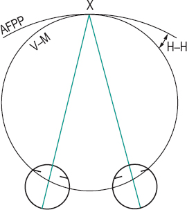
Hering–Hillebrand d. The deviation of the apparent frontoparallel plane horopter from the Vieth–Müller circle (horopter) (Fig. D2).
minimum d. of a prism See prism, minimum deviation of a.
primary d. The deviation found in paralysis of an extraocular muscle when the unaffected eye is fixating.
secondary d. The deviation found in paralysis of an extraocular muscle when the eye with the paralytic muscle is fixating.
skew d. A form of strabismus, typically vertical, that does not follow any standard or typical pattern and is usually difficult to quantify. It may be due to a midbrain disorder, multiple sclerosis or myasthenia gravis.
vertical d. 1. Type of ocular deviation found in strabismus in which the deviating eye is rotated upward with respect to the fixating eye. 2. Upward ocular deviation of an occluded eye in the cover test, as found in hyperphoria or hypophoria.
Devic’s disease See disease, Devic’s.
dexamethasone See antiinflammatory drug.
dextroclination Rotation of the upper pole of the vertical meridian of an eye to the subject’s right. Syn. dextrocycloduction; dextrotorsion.
dextrocycloversion Rotation of the upper poles of the vertical meridians of both eyes towards the subject’s right.
See laevocycloversion.
dextrodeorsumversion Movement of the eyes down and to the right.
dextroduction Rotation of one eye to the right. See duction.
dextrophoria A tendency of the visual axes of both eyes to deviate to the right, in the absence of a stimulus to fusion.
See heterophoria; laevophoria.
dextrotorsion See dextroclination.
dextroversion Movement of both eyes to the right.
See version.
diabetes A chronic disease that occurs when the pancreas does not produce enough insulin, or alternatively, when the body cannot effectively use the insulin it produces. Insulin is a hormone that regulates blood sugar. Hyperglycaemia, or raised blood sugar, is a common effect of uncontrolled diabetes and over time leads to serious damage to many of the body’s systems, especially the nerves and blood vessels (definition of the World Health Organization). The most common types of diabetes are: Type 1 diabetes, which is characterized by a lack of insulin production and therefore dependent upon insulin administration and Type 2 diabetes, which is characterized by an ineffective use of insulin. Type 1 diabetes is the most common type in young people whereas type 2 is the most common diabetes and affects primarily but not exclusively adults and it is largely the result of obesity and physical inactivity. The main complications in the eye are retinopathy, cataract, rubeosis iridis, ocular motor nerve palsies, xanthelasma and ptosis.
See accommodative insufficiency; anisocoria; glaucoma, neovascular; glaucoma, open-angle; hypoxia; myopia, lenticular; paralysis of the sixth nerve; paralysis of the third nerve; pupil, Adie’s; tritanopia; vitrectomy; vitreous detachment.
diabetic maculopathy; retinopathy See under the nouns.
diagnosis 1. Term that indicates the disease (e.g. pulmonary tuberculosis) or the refractive error (e.g. compound myopic astigmatism) that a person has. 2. The art of determining a disease or visual anomaly based on the signs, symptoms and tests.
diagnostic positions of gaze See positions of gaze, diagnostic.
dialysis, retinal See retinal dialysis.
diameter, total (TD) The linear measurement (usually specified in millimetres) of the maximum external dimension of a contact lens. It is equal to the back optic zone diameter (BOZD) plus twice the width of each of the back peripheral optic zones (if any) or twice the width of the edge in a spherical lens. Formerly, it was called overall size (OS).
See optic zone diameter; v gauge.
diaphragm 1. In optics, an aperture generally round and of variable diameter placed in a screen and used to limit the field of view of a lens or optical system (field stop). It also limits stray light (light stop). Syn. stop; aperture-stop. 2. In anatomy, a dividing membrane.
diascope A projector used to project transparent objects.
dibrompropamidine See antibiotic.
dichlorphenamide See carbonic anhydrase inhibitors.
dichoptic Viewing a separate and independent field by each eye, in binocular vision, as for example in a haploscope.
dichroism Property exhibited by certain transparent substances of producing two different colours depending upon the thickness of substance traversed, the directions of transmission of light and/or viewing, the concentration of the substance, etc. The most common example is that of crystals (e.g. tourmaline) that absorb unequally the ordinary and extraordinary rays.
See anisotropic; crystal, dichroic; pleochroism.
dichromat Person having dichromatism, i.e. a deuteranope, a protanope or a tritanope.
dichromatism A form of colour vision deficiency in which all colours can be matched by a mixture of only two primary colours. The spectrum appears as consisting of two colours separated by an achromatic area (the neutral point). There are several types of dichromatism: deuteranopia, protanopia and tritanopia. Syn. daltonism; dichromatopsia; dichromatic vision.
See colour vision, defective; pigment, visual.
diclofenac See antiinflammatory drug.
dicoria A condition in which there are two pupils in one iris. It may be congenital or the result of surgery or injury. Syn. diplocoria.
See polycoria.
differential threshold See threshold, differential.
diffraction Deviation of the direction of propagation of a beam of light, which occurs when the light passes the edge of an obstacle such as a diaphragm, the pupil of the eye or a spectacle frame. There are two consequences of this phenomenon. First, the image of a point source cannot be a point image but a diffraction pattern. This pattern depends upon the shape and size of the diaphragm as well as the wavelength of light. Second, a system of close, parallel and equidistant grooves, slits or lines ruled on a polished surface can produce a light spectrum by diffraction. This is called a diffraction grating.
See disc, Airy’s; fringes, diffraction; theory, Maurice’s.
diffractive contact lens See lens, contact.
diffuser A device used to scatter light. It can be a reflecting surface (e.g. matt paint) or a transmitting medium (e.g. ground glass).
diffusion 1. Scattering of light passing through a heterogeneous medium, or being reflected irregularly by a surface, such as a sandblasted opal glass surface. Diffusion by a perfectly diffusing surface occurs in accordance with Lambert’s cosine law. In this case, the luminance will be the same, regardless of the viewing direction. 2. The passive movement of ions or molecules through a medium or across a semi-permeable membrane (e.g. the ciliary epithelium) in response to a concentration gradient until equilibrium is reached. It is one of the three mechanisms that create aqueous humour.
See light, diffuse; reflection, diffuse; ultrafiltration.
diffusion circle See blur circle.
diisopropyl fluorophosphate (DFP) See anticholinesterase.
dilator pupillae muscle See muscle, dilator pupillae.
dioptre 1. A unit proposed by Monoyer to evaluate the refractive power of a lens or of an optical system. It is equal to the product of the refractive index in the image space and the reciprocal of the focal length in metres. (Symbol: D.) Thus a lens with a focal length (in air) of 1 m has a power of 1 D, one with a focal length of 1/2 m, has a power of 2 D, etc. 2. It is also incorrectly used to represent a unit of curvature, being equal to the reciprocal of the radius of curvature expressed in metres.
Table D3
Relationship between dioptres and focal length (in air)
| focal length | ||
| dioptre value | (cm) | (in) |
| 0.25 | 400 | 157 |
| 0.50 | 200 | 79 |
| 1.00 | 100 | 39 |
| 1.50 | 67 | 26 |
| 2.00 | 50 | 20 |
| 2.50 | 40 | 16 |
| 3.00 | 33.3 | 13 |
| 4.00 | 25 | 10 |
| 5.00 | 20 | 7.9 |
| 6.00 | 16.7 | 6.6 |
| 7.00 | 14.3 | 5.6 |
| 8.00 | 12.5 | 4.9 |
| 9.00 | 11.1 | 4.4 |
| 10.00 | 10 | 3.9 |
| 12.00 | 8.3 | 3.3 |
| 14.00 | 7.1 | 2.8 |
| 16.00 | 6.2 | 2.4 |
| 20.00 | 5.0 | 2.0 |
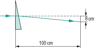
Table D4
Relationship between prism dioptres and degrees
| Prism dioptres (Δ) | degrees (°) | minutes (‘) |
| 1 | 0.573° | 0°34’ |
| 2 | 1.14° | 1°8’ |
| 3 | 1.71° | 1°43’ |
| 4 | 2.28° | 2°17’ |
| 5 | 2.85° | 2°51 ‘ |
| 6 | 3.43° | 3°26’ |
| 7 | 4.0° | 4°0’ |
| 8 | 4.57° | 4°34’ |
| 9 | 5.14° | 5°8’ |
| 10 | 5.71° | 5°43’ |
| 15 | 8.57° | 8°34 |
See curvature of a surface; myodioptre; paraxial equation, fundamental; refractive error; vergence.
dioptre, prism 1. A unit specifying the amount of light deviation by an ophthalmic prism. One prism dioptre (written 1 Δ) represents a deviation of 1cm on a flat surface 1 m away from the prism. The surface is perpendicular to the direction of the original light ray (Fig. D3). Similarly, a 2 Δ prism deviates light 2 cm at a distance of 1 m, and so on. For small angles, conversion between prism dioptres and degrees is given by the approximate formula

The exact formula for any angle α less than 90° is

Note: the current British Standard regarding ophthalmic lenses specifies a deviation (in Δ) of a ray of light of wavelength 587.6 nm incident normally at one surface. 2. A unit of convergence of the eyes.
See law, Prentice’s; power, prism.
dioptric power See power, refractive.
dioptrics That branch of optics which deals with the refraction of light (as opposed to reflection). Example: the dioptrics of the eye.
dipivefrine hydrochloride See sympathomimetic drugs.
diplocoria See dicoria.
diplopia The condition in which a single object is seen as two rather than one. This is usually due to images not stimulating corresponding retinal areas. Other causes are given below. Syn. double vision (colloquial).
See effect, differential prismatic; haplopia; myasthenia gravis; retinal corresponding points; polyopia; sclerosis, multiple; strabismus; test, diplopia; triplopia.
binocular d. Diplopia in which one image is seen by one eye and the other image is seen by the other eye.
crossed d. See diplopia, heteronymous.
heteronymous d. Binocular diplopia in which the image received by the right eye appears to the left and that received by the left eye appears to the right. In this condition the images are formed on the temporal retina. Syn. crossed diplopia.
homonymous d. Binocular diplopia in which the image received by the right eye appears to the right and that received by the left eye appears to the left. In this condition, the images are formed on the nasal retina. Syn. uncrossed diplopia.
incongruous d. Diplopia present in individuals with abnormal retinal correspondence in which the relative positions of the two images differ from what would be expected on the basis of normal retinal correspondence. Example: an exotrope experiencing homonymous diplopia instead of heteronymous diplopia. Syn. paradoxical diplopia.
See retinal correspondence, abnormal.
monocular d. Diplopia seen by one eye only. It is usually caused by irregular refraction in one eye (e.g. in early cataracts, corneal opacity) or by dicoria or polycoria. It may be induced by placing a biprism in front of one eye.
See image, ghost; luxation of the lens.
paradoxical d. See diplopia, incongruous.
pathological d. Any diplopia due to an eye disease (e.g. proptosis), an anomaly of binocular vision (e.g. strabismus), a variation in the refractive index of the media of the eye (e.g. cataract), a subluxation of the crystalline lens, or to a general disease (e.g. multiple sclerosis, myasthenia gravis).
See exophthalmos; luxation of the lens.
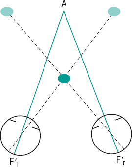
physiological d. Normal phenomenon which occurs in binocular vision for non-fixated objects whose images fall on disparate retinal points. It is easily demonstrated to persons with normal binocular vision: fixate binocularly a distant object and place a pencil vertically some 25 cm in front of your nose. You should see two rather blurred pencils. The observation of physiological diplopia has been found to be useful in the management of eso or exo deviations, suppression, abnormal retinal correspondence, etc. (Fig. D4).
See Brock string; disparity, retinal.
uncrossed d. See diplopia, homonymous.
diploscope Instrument used to evaluate binocular vision and which may be used for the treatment of anomalies of binocular vision.
direct ophthalmoscopy See ophthalmoscopy, direct.
direction, oculocentric Direction associated with a particular retinal point. It is always perceived in the same direction if the light is received by the same retinal receptor. The capacity of a receptor to distinguish its excitation from that of its neighbours is referred to as local sign (or Lotze’s local sign). This characteristic means that each retinal receptor has a unique oculocentric direction.
See line of direction; oculocentre.
disc 1. A flat, circular, coin-shaped structure. 2. In anatomy, the intervertebral disc.
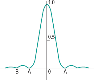
Airy’s d. Owing to the wave nature of light, the image of a point source consists of a diffraction pattern. If light passes through a circular aperture, the diffraction pattern will appear as a bright central disc, called Airy’s disc, surrounded by concentric light and dark rings. Airy’s disc receives about 87% of the luminous flux, the next concentric ring about 8%, and the next 3%. The radius of Airy’s disc equals

where d is the radius of the entrance pupil of the optical system of focal length f and λ the wavelength of the light used. In the eye, with a pupil of 4 mm diameter and λ = 507 nm, the diameter of Airy’s disc is about 5 mm, which corresponds to a visual angle of about one minute of arc (Fig. D5). Syn. diffraction disc.
See criterion, Rayleigh; function, point-spread; resolution, limit of.
choked d. See papilloedema.
cupped d. An enlarged and deepened excavation of the physiological cup. It may be physiological, or due to glaucoma (glaucomatous cup), or following atrophy of the optic nerve (as in papilloedema).
diffraction d. See disc, Airy’s.
Maxwell d. A rotating disc onto which differently coloured discs which are radially slit can be fitted together to overlap and divide the surface into sectors of different colours. It may be used to investigate colour mixture.
morning glory d. A congenital, usually unilateral, anomaly of the optic disc. It may be due to a failure of the embryonic fissure such that the optic disc and some peripapillary tissue prolapse posteriorly. The optic disc is abnormally large and a white-grey tuft of glial tissue covers its centre. The annular zone surrounding the disc has irregular areas of pigmentation and depigmentation. The optic disc thus resembles a morning glory flower. Patients present with reduced visual acuity and strabismus and, in about one-third of patients, retinal detachment.
optic d. Region of the fundus of the eye corresponding to the optic nerve head. It can be seen with the ophthalmoscope as a pinkish-yellow area with usually a whitish depression called the physiological cup. The optic disc has an area of about 2.7 mm2, a horizontal width of about 1.75 mm and a vertical height of about 1.9 mm. The optic disc is the anatomical correlate of the physiological blind spot. It is greatly affected in glaucoma, papillitis, Leber’s hereditary optic atrophy. Syn. optic nerve head; optic papilla (this is not strictly correct because the disc is not elevated above the surrounding retina).
See cup, glaucomatous; drusen, optic disc; neuroretinal rim; papilloedema; syndrome, Swann’s.
pinhole d. (ph) A blank disc with a small aperture (2 mm diameter or less) mounted in a trial lens rim. It is used to reduce the size of the blur circle in an ametropic eye. In this condition vision will improve giving an indication of the final visual acuity that will be obtained with corrective lenses. If no improvement occurs, the eye is amblyopic. This procedure is called the pinhole test.
Scheiner’s d. An opaque disc in which there are two pinholes separated by a distance less than the pupil diameter. It is used to measure the dioptric changes during accommodation or to detect the type of ametropia (Fig. D6).
See experiment, Scheiner’s.
situs inversus of the d. A congenital, usually bilateral condition in which the retinal vessels course nasally from the disc instead of temporally. It is often associated with congenital scleral crescent and myopic astigmatism.
stenopaeic d. 1. A pinhole disc. 2. A blank disc with a slit used in detecting and measuring the astigmatism of the eye (Fig. D7).
Syn. stenopaeic slit. Note: also spelt stenopeic or stenopaic.
See kinescope; spectacles, stenopaeic.
tilted d. A congenital, bilateral condition in which the optic nerves insert obliquely into the globe. It is often associated with congenital scleral crescent and myopic astigmatism. The only sign is a bitemporal visual field defect (often upper temporal).
disciform keratitis See keratitis, disciform.
disciform scar A subretinal scar, most often located in the macular area. It results from the haemorrhages that sometimes follow choroidal neovascularization (consisting of fibrovascular tissue) in the exudative type of age-related macular degeneration. It causes irreparable damage to vision.
discomfort glare See glare, discomfort.
disconjugate movements See movements, disjunctive eye.
disease An abnormal process affecting the structure or function of a part, organ or system of the body. It is typically manifested by signs and symptoms, but the aetiology may or may not be known. Disease is a response to a specific infective agent (a microorganism or a poison), to environmental factors (e.g. malnutrition, injury, industrial hazards), to congenital or hereditary defects, or to a combination of all these factors. Note: illness is sometimes used as a synonym of disease, but it also refers to a person’s perception of their health, regardless of whether the person does or does not have a disease.
autoimmune d. A disease produced when the immune response of an individual is directed against its own cells or tissues. It is not yet known exactly what causes the body to react to one’s own antigens as if they were foreign. Examples: diabetes mellitus type 1; Graves’ disease; multiple sclerosis; myasthenia gravis; rheumatoid arthritis; Reiter’s disease; Sjögren’s syndrome.
Batten–Mayou d. Juvenile form of amaurotic family idiocy. It is characterized by progressive degeneration of the retina, which eventually leads to blindness. Syn. Spielmeyer–Stock disease.
Behçet’s d. See syndrome, Behçet’s.
Benson’s d. See asteroid hyalosis.
Berlin’s d. A traumatic phenomenon in which the posterior pole of the retina develops oedema (and haemorrhages). Syn. commotio retinae.
Best’s d. An autosomal dominant inherited degeneration in which there is an accumulation of lipofuscin within the retinal pigment epithelium, which interferes with its function. It is caused by a mutation in bestrophin gene (BEST1). The disease is characterized by the appearance on the retina in the first and second decades of life of a bright orange deposit, resembling the yolk of an egg (vitelliform), with practically no effect on vision. It eventually absorbs, leaving scarring, pigmentary changes and impairment of central vision in most cases, although in some cases the retinal lesion may be eccentric, with very little effect on vision. The electrooculogram is abnormal throughout the development of the disease from pre-vitelliform, vitelliform and the end-stage when there is scarring or atrophy. Syn. Best’s macular dystrophy; juvenile vitelliform macular dystrophy; vitelliform macular dystrophy. Mutation in the VMD2 gene can cause adult vitelliform macular dystrophy, a condition characterized by smaller macular lesions and very little impairment of vision.
Bowen’s d. A disease characterized by a slow-growing tumour of the epidermis of the skin which may involve the corneal or conjunctival epithelium.
Coats’ d. Chronic, progressive retinal vascular anomalies, usually unilateral, occurring predominantly in young males. It is characterized by retinal exudates, irregular dilatation (telangiectasia) and tortuosity of retinal vessels and appears as a whitish fundus reflex (leukocoria). Subretinal haemorrhages are frequent and eventually retinal detachment may occur. The main symptom is a decrease in central or peripheral vision, although it may be asymptomatic in some patients. Management may involve photocoagulation or cryotherapy. A less severe form of the disease is called Leber’s miliary aneurysms. Syn. retinal telangiectasia.
Crohn’s d. A type of inflammatory, chronic bowel disease characterized by granulomatous inflammation of the bowel wall causing fever, diarrhoea, abdominal pain and weight loss. The ocular manifestations include acute iridocyclitis, scleritis, conjunctivitis and corneal infiltrates.
Devic’s d. A demyelinative disease of the optic nerve, the optic chiasma and the spinal cord characterized by a bilateral acute optic neuritis with a transverse inflammation of the spinal cord. Loss of visual acuity occurs very rapidly and is accompanied by ascending paralysis. There is no treatment for this disease. Syn. neuromyelitis optica.
Eales’ d. A non-specific peripheral retinal periphlebitis (i.e. an inflammation of the outer coat of a vein) that usually affects mostly young males, often those who have active or healed tuberculosis. It is characterized by recurrent haemorrhages in the retina and vitreous. This disease is a prime example of retinal vasculitis.
Fabry’s d. An X-linked recessive disease caused by mutations in the gene encoding alpha-galactosidase A (GLA) and characterized by an abnormal accumulation of glycolipid in the tissues. It appears as small purple skin lesions on the trunk and there may be renal and cardiovascular abnormalities. Ocular signs include whorl-like corneal opacities, star-shaped lens opacities, and tortuous conjunctival and retinal blood vessels.
Graves’ d. An autoimmune disorder in which immunoglobulin antibodies bind to thyroid-stimulating hormone receptors in the thyroid gland and stimulate secretion of thyroid hormones leading to hyperthyroidism. The main ocular manifestations (called Graves’ ophthalmopathy) are exophthalmos, retraction of the eyelids (Dalrymple’s sign), conjunctival hyperaemia, lid lag in which the upper lid follows after a latent period when the eye looks downward (von Graefe’s sign), defective eye movements (restrictive myopathy) and optic neuropathy, besides increased pulse rate, tremors, loss of weight and diarrhoea. It typically affects women between the ages of 20 and 50 years. Most common signs associated with the disease are those of von Graefe and Moebius. Syn. thyrotoxicosis. If only the eye signs of the disease are present without clinical evidence of hyperthyroidism, the disease is called euthyroid or ophthalmic Graves’ disease. Treatment begins with control of the hyperthyroidism (if present). Some cases may recover spontaneously with time. Mild cases of ocular deviations and restrictions may benefit from a prismatic correction. Corticosteroids and radiotherapy may be needed and surgery is a common form of management, especially when there is diplopia in the primary position of gaze.
See accommodative infacility; exophthalmos; ophthalmopathy, thyroid.
Harada’s d. A disease characterized by bilateral exudative uveitis associated with alopecia, vitiligo and hearing defects. However, as many aspects of this entity overlap clinically and histopathologically with the Vogt–Koyanagi syndrome it is nowadays combined and called the Vogt– Koyanagi–Harada syndrome.
von Hippel’s d. A rare disease, sometimes familial, in which haemangiomata occur in the retina where they appear ophthalmoscopically as one or more round, elevated reddish nodules. The condition is progressive and takes years before there is a complete loss of vision. Syn. angiomatosis retinae.
von Hippel–Lindau d. Retinal haemangioblastoma involving one or both eyes associated with similar tumours in the cerebellum and spinal cord and sometimes cysts of the kidney and pancreas. Ophthalmoscopic examination shows a reddish, slightly elevated tumour.
Leber’s d. See Leber’s hereditary optic atrophy.
Niemann–Pick d. An autosomal recessive inherited lipid storage disorder characterized by a partial destruction of the retinal ganglion cells and a demyelination of many parts of the nervous system. It is caused by mutation in the NPC1 gene. The condition usually involves children of Jewish parentage. When the retina is involved, there is a reddish central area (cherry-red spot) surrounded by a white oedematous area. The disease usually leads to death by the age of two. This disease is differentiated from Tay–Sachs disease because of its widespread involvement and gross enlargement of the liver and the spleen. Syn. sphingomyelin lipidosis.
Norrie’s d. An inherited X-linked recessive disorder characterized by bilateral congenital blindness. It is caused by mutation in the norrin gene (NDP). The initial ocular presentation is leukocoria. It then progresses to cataract, corneal opacification and phthisis bulbi. The condition may be associated with mental retardation and hearing defects. Syn. oculoacoustico-cerebral degeneration; Andersen–Warburg syndrome.
Oguchi’s d. An autosomal recessive, inherited night blindness occurring mainly in Japan. All other visual capabilities are usually unimpaired but the patient presents an abnormal golden brown fundus reflex in the light-adapted state, which becomes a normal colour with dark-adaptation (Mizuo phenomenon). It is presumed to be due to an abnormality in the neural network of the retina. The disease can be caused by mutation in the arrestin gene (SAG) or the rhodopsin kinase gene (GRK1).
ophthalmic Graves’ d. See disease, Graves’.
Paget’s d. Hereditary systemic disorder of the skeletal system accompanied by visual disturbances, the most common being retinal arteriosclerosis.
See angioid streaks; arteriosclerosis.
von Recklinghausen’s d. An autosomal dominant inherited disease with a gene locus at 17q11. It is caused by mutation in the neurofibromin gene. It is characterized by tumours in the central nervous system and in cranial nerves, enlarged head, ‘café au lait’ spots on the skin, choroidal naevi, optic nerve glioma, peripheral neurofibromas (e.g. on the eyelid) and Lisch nodules. Glaucoma may occur. Syn. neurofibromatosis type 1 (NF-1).
Refsum’s d. See syndrome, Refsum’s.
Reiter’s d. A systemic syndrome characterized by a triad of three diseases: urethritis, arthritis and conjunctivitis. Keratitis and iridocyclitis may follow as complications. It occurs mainly in young men typically following urethritis and less commonly after an attack of dysentery or acute arthritis, which usually affects the knees, ankles and Achilles tendon. Syn. Reiter’s syndrome.
Sandhoff’s d. An autosomal recessive inherited disease similar to Tay–Sachs disease with the same signs, but differing in that both the enzymes hexosaminidase A and B are defective and it develops more rapidly and can be found among the general population. The main ocular manifestation is a whitish area in the central retina with a cherry-red spot which eventually fades and the optic disc develops atrophy. Syn. Gm2 gangliosidosis type2.
sickle-cell d. A hereditary anaemia encountered among black and dark-skinned people due to a defect in the haemoglobin. It is characterized by retinal neovascularization, haemorrhages and exudates, cataract and subconjunctival haemorrhage. Syn. sickle-cell anaemia.
Spielmeyer–Stock d. See disease, Batten–Mayou.
Stargardt’s d. An autosomal recessive inherited disorder of the retina occurring in the first or second decade of life and affecting the central region of the retina. A few cases are inherited as an autosomal dominant trait. Known causes of the disease include a mutation in one of the following genes: ABCA4, CNGB3 and ELOVL4. There is an accumulation of lipofuscin within the retinal pigment epithelium, which interferes with its function. With time a lesion develops at the macula, which has a ‘beaten-bronze’ reflex. It is often surrounded by yellow-white flecks. There is a loss of central vision but peripheral vision is usually normal. Myopia is very common. Management usually consists of a high plus correction for near to magnify the retinal image and wearing UV-protecting sunglasses. Syn. Stargardt’s macular dystrophy.
See dysrophy, macular; fundus flavimaculatus .
Steinert’s d. See dystrophy, myotonic.
Still’s d. See arthritis, juvenile rheumatoid.
Sturge–Weber d. See syndrome, Sturge–Weber.
Tay–Sachs d. An autosomal recessive lipid storage disorder caused by a deficiency of the enzyme hexosaminidase A which leads to an accumulation of Gm2 ganglioside (a fatty acid derivative) in the ganglion cells of both the retina and the brain. It has its onset in the first year of life, vision is affected and the central retina shows a whitish area with a reddish central area (cherry-red spot), which fades and the optic disc develops atrophy. Eventually the eye becomes blind and death occurs, usually at about the age of 30 months. It affects Jewish infants more than others by a factor of about ten to one. Syn. Gm2 gangliosidosis type 1; infantile amaurotic familial idiocy.
See disease, Niemann–Pick.
Terrien’s d. See ectasia, corneal.
Wagner’s d. See syndrome, Wagner’s.
Wernicke’s d. A disease characterized by disturbances in ocular motility, pupillary reactions, nystagmus and ataxia. It is mainly due to thiamin deficiency and is frequently encountered in chronic alcoholics. Syn. Wernicke’s syndrome.
Wilson’s d. A systemic disease resulting from a deficiency of the alpha-2-globulin ceruloplasmin beginning in the first or second decade of life. It is characterized by widespread deposition of copper in the tissues, tremor, muscular rigidity, irregular involuntary movements, emotional instability and hepatic disorders. The ocular features are degenerative changes in the lenticular nucleus and most noticeably a Kayser–Fleischer ring. Syn. hepatolenticular degeneration; lenticular progressive degeneration; pseudosclerosis of Westphal.
disinfectant See antiseptic.
disinfection The process or act of destroying pathogenic microorganisms. However, certain bacterial spores may survive and germinate which could lead to contamination.
See antiseptic; sterilization; surfactant.
chemical d. A method of disinfecting soft contact lenses, using solutions containing either a preservative or hydrogen peroxide. Preservatives include chlorhexidine, thimerosal (very rarely used nowadays) and more commonly nowadays the preservatives with larger molecules which cannot penetrate into the lens matrix of soft contact lenses, such as the biguanide polyhexanide (polyaminopropyl biguanide or polyhexamethylene biguanide). Hydrogen peroxide has a broad-spectrum efficacy against bacteria, fungi and viruses. It must, however, be neutralized before the lens can be worn. Rigid gas permeable contact lenses are disinfected with a preservative such as chlorhexidine, benzalkonium chloride, polyhexanide and polixetonium chloride. Failure to disinfect contact lenses may lead to microbial keratitis. Disinfectants for contact lenses have to pass FDA and International Organization for Standardization (ISO) tests to be approved. They must be effective against three specific bacteria (Pseudomonas aeruginosa, Staphylococcus aureus and Serratia marcescens) and two fungi (Candida albicans and Fusarium solani).
See neutralization.
heat d. A method of disinfecting soft contact lenses, based on heating the lens to a temperature of at least 80° for 10 minutes. This is achieved in specially manufactured heating units in which the lenses are kept in physiological saline solution. However, repeated boiling of soft lenses may cause some degradation of the lens material, and tear mucoproteins that have not been previously removed with a surface cleaning agent tend to become coagulated on the lens surface.
disintersion, retinal See retinal dialysis.
disjunctive movements; nystagmus See under the nouns.
dislocation of the lens See luxation of the lens.
disparate retinal points Non-corresponding retinal points.
disparity The condition of being unequal or totally different. The word is used mainly to refer to non-corresponding points in the retina.
See disparity, retinal; retinal corresponding points.
binocular d. See acuity, stereoscopic visual; disparity, retinal; perception, depth.
crossed d. Retinal disparity induced by an object nearer to the eyes than the point of fixation and focused on the temporal retina. Thus, the image received by the right eye appears to the left and that received by the left eye appears to the right. Syn. crossed retinal disparity.
fixation d. See disparity, retinal.
retinal d. Binocular vision in which the two retinal images of a single object do not fall on corresponding retinal points, i.e. when the object lies off the horopter. If, however, the two retinal images still fall within Panum’s area, the object will still be seen single. At the fixation point this may cause over- or under-convergence of the eyes. This particular case is called fixation disparity (or retinal slip). The presence of fixation disparity often indicates that binocular vision is under stress and the patient has an uncompensated heterophoria. Optical correction or orthoptic exercises usually eliminate the symptoms. Fixation disparity can be measured either (1) directly (e.g. Disparometer, Wesson Fixation Disparity Card, both consisting of targets with pairs of vernier lines of various angular separation, each line being seen by one eye through polarizing filters), or (2) indirectly as an associated phoria (e.g. Mallett fixation disparity unit). Syn. binocular disparity.
See acuity, stereoscopic visual; anaglyph; esodisparity; exodisparity; fusional movements; perception, depth; stereogram, random-dot; stereopsis; stereotest.
uncrossed d. Retinal disparity induced by an object farther away from the eyes than the point of fixation and focused on the nasal retina. Thus, the image received by the right eye appears to the right and that received by the left eye appears to the left. Syn. uncrossed retinal disparity.
vergence d. See fusion, motor.
Disparometer Trade name for a clinical instrument designed to measure fixation disparity at near. The target does not have a binocular fixation point and the fusion lock is parafoveal. The instrument fits on the near point rod of a standard phoropter. The Disparometer has two stimuli: one for vertical disparity measurement and the other for horizontal disparity measurement. The test consists of successive pairs of vernier lines of increasing angular separation within a structureless field, each line being viewed by one eye through polarizing filters. The edge of the field provides a peripheral fusion stimulus. Fixation disparity is measured when the vernier lines appear to be aligned and the amount is given by the angular separation (in minutes of arc) of the lines indicated on the back of the instrument. A fixation disparity curve can be obtained by determining the fixation disparity for various amounts of prism power placed in front of the eyes.
See heterophoria, associated; Mallett fixation disparity unit.
dispensing, optical The act of issuing an optical appliance which corrects, remedies or relieves defects of vision (definition of the World Council of Optometry). Syn. dispensing; ophthalmic dispensing.
See optician, dispensing; optics, ophthalmic.
dispersion Phenomenon of the change in velocity of propagation of radiation in a medium, as a function of its frequency, which causes a separation of the monochromatic components of a complex radiation. All optical media cause dispersion by virtue of their variation of refractive index with wavelengths. Dispersion is specified by the difference in the refractive index of the medium for two wavelengths. The difference between the blue F (486.1 nm) and the red C (656.3 nm) spectral lines is called the mean dispersion, i.e. nF – nC. Dispersion is usually represented by its dispersive power ω or relative dispersion which is equal to the mean dispersion divided by the excess refractive index of the helium d (587.6 nm) spectral line (nd – 1), often called the refractivity of the material,

The reciprocal of the dispersive power is called the Abbé’s number or constringence (Fig. D8).
See aberration longitudinal chromatic; axis, achromatic; lines, Fraunhoffer’s; prism, achromatic.
dispersion, mean; relative See dispersion.
dispersive power See dispersion.
disposable contact lens See lens, contact.
dissociated nystagmus See nystagmus.
dissociated vertical deviation See deviation, dissociated vertical.
dissociated vertical divergence See hyperopia, alternating.
dissociating test See test, dissociating.
dissociation Elimination of the stimulus to fusion. It is usually accomplished by occluding one eye, or by inducing gross distortion of the image seen by one eye (e.g. Maddox rod), or by placing a strong prism in front of one eye (e.g. von Graefe’s test) with the result that the eyes will move to the passive position (or heterophoria position).
See heterophoria, dissociated; position, passive; test, diplopia; test, dissociating.
distal Farthest from a central point.
See proximal.
distance The amount of space between two points.
abathic d. See plane, apparent frontoparallel.
d. between lenses (DBL) Horizontal distance between the nasal parts of the spectacle lenses in a frame, measured either along the datum line (datum system) or between the nasal peaks of the bevels of the two spectacle lenses (boxing system).
See spectacle frame markings; system, boxing.
d. between rims (DBR) Horizontal distance between the bearing surfaces of a regular bridge of a spectacle frame, usually measured along the datum line, or at a specified distance below the crest of the bridge.
centration d. (CD) The specified horizontal distance between the right and left centration points of a pair of ophthalmic lenses.
See distance, near centration; point, centration.
conjugate d’s. An optical system will form an image of an object. As the path of light is reversible, the position of object and image are interchangeable. These pairs of object and image points are called conjugate points (or conjugate foci) and the distances of the object and the image from the optical surface are called the conjugate distances (Fig. D9). When an eye is accurately focused for an object, object and retina are conjugate.
See emmetropia; experiment, Scheiner’s.
d. of distinct vision A conventional distance used in calculating the magnifying power of a loupe or microscope. It is usually taken as 25 cm (or 10 inches) from the eye.
See magnification, apparent.
hyperfocal d. That distance from a lens or optical system at which the depth of field, on the far side of an object in focus, extends to infinity. On the near side of the object the depth of field then extends to half that distance. This is a useful distance in photography as it represents the shortest distance on which to focus in order to obtain a reasonable image definition of an object at infinity and the longest total depth of field. This distance depends on the focal length and the diameter of the entrance pupil of the system as well as the amount of the allowable blur.
See depth of field.
image d. The distance along the optical axis of a lens or optical system between the image plane and the secondary principal plane. If the system consists of a single thin lens, the image distance is measured from the optical surface and the reciprocal of this quantity is called the reduced image vergence or image vergence (in air).
See plane, principal; power, back vertex.
interocular d. The distance between the centres of rotation of the eyes, i.e. the length of the base line.
interpupillary d. (IPD, PD) The distance between the centres of the pupils of the eyes. It usually refers to the eyes fixating at distance, otherwise reference must be made to the fixation distance (e.g. near interpupillary distance). The average interpupillary distance for men is about 65 mm and for women about 62 mm (in Caucasians). Syn. pupillary distance. The interpupillary distance is often measured from the median plane to the centre of the pupil of each eye. This is referred to as the monocular pupillary distance (MPD): it is a useful measurement, especially in dispensing progressive lenses. The interpupillary distance for near vision can be calculated using the following formula:

where d is the distance between the target plane and the spectacle plane and d’ the distance between the target plane and the midpoint between the centres of rotation of the eyes.
See pupillometer; rule, PD.
near centration d. (NCD) The horizontal distance between the right and left centration points used for near vision.
object d. The distance along the optical axis of a lens or optical system between the object plane and the primary principal plane. If the system consists of a single thin lens, the object distance is measured from the optical surface and the reciprocal of this quantity is called the reduced object vergence or object vergence (in air).
See power, front vertex; vergence.
reading d. The normal distance at which people read. It is about 33–44 cm for men and 29–40 cm for women. It is a useful measurement in determining the reading addition.
vertex d. See vertex distance.
d. vision See vision, distance.
working d. (WD) 1. The distance at which a person reads or does close work. 2. In retinoscopy, the distance between the plane of the sighthole and that of the patient’s spectacles. 3. In microscopy, the distance between an object and the front surface of the objective.
distichiasis Congenital anomaly in which there is a double row of eyelashes in the lid margin, one row being normal and the other row turning inward towards the eye. Distichiasis can also be acquired following scarring or chemical and physical injury. If there are symptoms, treatment consists of removal of the aberrant eyelashes, usually by cryotherapy (removal under cold or freezing conditions) or electrolysis.
See epilation; polystichia; trichiasis.
distometer An instrument for measuring the distance between the back surface of a spectacle lens and the apex of the cornea. It is usually carried out by having the patient close the eyes. One end of the instrument or caliper rests against the upper eyelid and the other presses against the back surface of the spectacle lens. The measurements are most commonly given to the nearest 0.5 mm. Syn. Lenscorometer (a tradename); vertexometer.
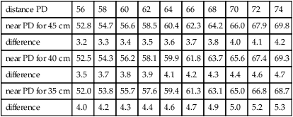
Table D5
Calculated near PD (in mm) as a function of distance PD for three reading distances (target plane to spectacle plane). The distance between the spectacle plane and the midpoint of the base line is assumed to be 27 mm (vertex distance 12 mm)
| distance PD | 56 | 58 | 60 | 62 | 64 | 66 | 68 | 70 | 72 | 74 |
| near PD for 45 cm | 52.8 | 54.7 | 56.6 | 58.5 | 60.4 | 62.3 | 64.2 | 66.0 | 67.9 | 69.8 |
| difference | 3.2 | 3.3 | 3.4 | 3.5 | 3.6 | 3.7 | 3.8 | 4.0 | 4.1 | 4.2 |
| near PD for 40 cm | 52.5 | 54.3 | 56.2 | 58.1 | 59.9 | 61.8 | 63.7 | 65.6 | 67.4 | 69.3 |
| difference | 3.5 | 3.7 | 3.8 | 3.9 | 4.1 | 4.2 | 4.3 | 4.4 | 4.6 | 4.7 |
| near PD for 35 cm | 52.0 | 53.8 | 55.7 | 57.6 | 59.4 | 61.3 | 63.1 | 65.0 | 66.8 | 68.7 |
| difference | 4.0 | 4.2 | 4.3 | 4.4 | 4.6 | 4.7 | 4.9 | 5.0 | 5.2 | 5.3 |
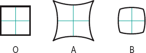
distortion Aberration of an optical system resulting in an image which does not conform to the shape of the object, somewhat resembling the image viewed through a cylindrical lens. This is due to an unequal magnification of the image. Distortion can be barrel-shaped (barrel-shaped distortion) in which the corners of the image of a square are closer to the centre than the middle part of the sides; or pincushion (pincushion distortion) in which the corners of the image of a square are farther from the centre than the middle part of the sides (Fig. D10). Example of barrel-shaped distortion: a square object seen through an uncorrected negative spectacle lens. Example of pincushion distortion: a square object seen through an uncorrected positive spectacle lens.
See correction; lens, fisheye; sine condition.
distortion test See test, distortion.
diurnal cycle See rhythm, circadian.
diurnal variations, in intraocular pressure Normal intraocular pressure varies throughout the day within a range of about 4 mmHg, being higher in the morning than in the evening. In patients with primary open-angle glaucoma this range is greater. This variation must be taken into consideration when measuring intraocular pressure.
diurnal vision See vision, diurnal.
divergence 1. Movement of the eyes turning away from each other. 2. Characteristic of a pencil of light rays, as when emanating from a point source. Syn. negative convergence.
See vergence.
d. excess A high exophoria at distance associated with a much lower exophoria at near. It may occasionally give rise to diplopia in distance vision.
fusional d. A movement of the eyes away from each other in response to retinal disparity, in order to restore single binocular vision. It occurs most commonly when induced by a base-in prism.
d. insufficiency A high esophoria at distance associated with esophoria at near. It often gives rise to symptoms of asthenopia in both distance and near vision.
d. paralysis See paralysis, divergence.
vertical d. Relative vertical movement between the two eyes.
diverging lens See lens, diverging.
DNA (deoxyribonucleic acid) A type of nucleic acid that constitutes the molecular basis of heredity. It is found principally in the nucleus of all cells where it forms part of the chromosome, or in the cytoplasm of cells lacking a nucleus, such as bacteria. It acts as the carrier of genetic information containing the instructions (code) to make proteins. It consists of two single chains of nucleotides, which are twisted round each other to form a double helix or spiral. The nucleotides contain sugar (deoxyribose), phosphate and the bases (adenine, cytosine, guanine and thymine). The two strands of DNA are held together by hydrogen bonds located between specific pairs of bases (adenine to thymine and cytosine to guanine). The sequence of bases and consequently gene sequence is sometimes altered, causing mutation. Assessment of DNA has found many applications, including forensic science to help identify a perpetrator (a process called genetic fingerprinting), to establish family relationships or the history of a particular population (phylogenetics).
See chromosome; gene; inheritance; mutation.
doll’s head manoeuvre; phenomenon
See phenomenon, doll’s head.
Dolman’s test See test, hole in the card.
dominance, ocular The superiority of one eye whose visual function predominates over the other eye. It is that eye (called the dominant eye) which is relied upon more than the other in binocular vision. It is not necessarily the eye with the best acuity. The lack of ocular dominance is referred to as ambiocularity and such a person is ambiocular.
See manoptoscope; occlusion treatment; test, hole in the card.
dominance, ocular column See column, cortical.
dominant eye; inheritance See under the nouns.
dominant wavelength (of a colour stimulus, not purple) See wavelength, dominant.
Donders’ diagram Graphical representation of total convergence as a function of accommodation for any fixation distance. The accommodation in dioptres is represented on the ordinate and the convergence in prism dioptres (or metre angles) on the abscissa. It is used to represent the binocular status of the two eyes, as well as evaluating the patient’s visual discomfort at any distance.
See accommodation, relative amplitude of; convergence, relative; line, demand; zone of clear, single, binocular vision.
Donders’ law See law, Donders’.
Donders’ line See line, demand.
Donders’ method See method, push-up.
Donders’ reduced eye See eye, reduced.
Doppler’s ophthalmodynanometer See ophthalmodynanometer.
dorsal Relating to either the back (posterior), or to the top in brain orientation.
See system, magnocellular visual; ventral.
dorsal midbrain syndrome See syndrome, Parinaud’s.
dorzolamide See carbonic anhydrase inhibitors.
dot haemorrhage See haemorrhage, blot.
double elevator palsy See palsy, double elevator.
double lid eversion See eversion, lid.
double refraction See birefringence.
double prism test See test, double prism.
double vision See diplopia.
doublet A combination of two lenses usually cemented to each other used to correct chromatic aberration. Typically it consists of a positive crown lens and a negative flint lens.
See lens, achromatizing; triplet.
Down’s syndrome See syndrome, Down’s.
downbeat nystagmus See nystagmus.
Doyne’s honeycomb choroiditis See drusen, familial dominant.
Draper’s law See law, Draper’s.
drift See movements, fixation.
droopy eyelid See ptosis.
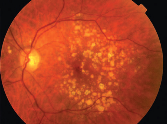
drusen Small, circular, yellow or white dots located throughout the fundus but more so in the macular region, around the optic disc or the periphery. They consist of deposits of abnormal extracellular material (amyloid P, complement proteins (C3, C5, C5b-9 complex), factors C, apolipoproteins B and E, lipids, vitronectin, etc.) derived mainly from the retinal pigment epithelium (RPE) and neural retina and they are located between the basement membrane of the RPE and Bruch’s membrane. Drusen interfere with the blood supply to the photoreceptors. Although they may be found in young people, they almost universally occur with ageing but also with retinal and choroidal degeneration (e.g. age-related maculopathy, retinitis pigmentosa, angioid streaks) and primary dystrophy (e.g. fundus flavimaculatus). There are several main types of drusen: (1) Hard (or nodular) drusen are small, round and discrete. They are deposits of granular material as well as of abnormal collagen. They are the most common type and are usually innocuous. (2) Soft (or diffuse or granular) drusen are often large with indistinct edges and with time they may enlarge, coalesce and increase in number. They are due to either a focal thickening of the inner layer of Bruch’s membrane or to amorphous material located between that thickened, detached part and the rest of Bruch’s membrane. They represent an early feature of age-related macular degeneration. (3) Cuticular (or basal laminar) drusen are small subretinal nodular thickening of the basement membrane of the pigment epithelium. They occur in younger patients more often than hard or soft drusen. (4) With time, the above drusen may calcify (calcific drusen) and take on a glistening appearance. Drusen rarely produce any symptoms and if there is a visual loss it is usually due to an accompanying macular haemorrhage, but if the drusen are very large thus widening the separation between the RPE and Bruch’s membrane there may be a degeneration of the overlying RPE and photoreceptors (Fig. D11). Syn. colloid bodies; hyaline bodies.
See macular degeneration, age-related; naevus, choroidal.
familial dominant d. An autosomal dominant hereditary degeneration of the choroid characterized by light-coloured patches of colloid material in the area around the macula and often the optic disc. The majority of cases are caused by mutations in the EFEMP1 gene (egf-containing fibulin-like extracellular matrix protein 1). There is no loss of vision unless it is followed by macular degeneration. Syn. Doyne’s honeycomb choroiditis; Doyne’s honeycombed degeneration; Tay’s choroiditis (used more commonly for the elderly).
optic disc d. Whitish-yellow spherical excrescences that lie on, or within, or occasionally around the optic nerve head. They are composed of calcified hyalinelike material possibly resulting from deposition of mucoprotein and calcium that have extruded from degenerating axons. In childhood they are usually buried within the disc substance and thus not visible on clinical examination but cause elevation of the disc surface resembling papilloedema. With age they become progressively more superficial. Field defects are common (e.g. generalized constriction, blind spot enlargement) but visual acuity is normal, unless there is some vascular complication. They usually appear bilaterally and affect males and females equally. They are easily diagnosed with fluorescein angiography because exposed drusen are autofluorescent.
dry eye See eye, dry; glands, meibomian; keratoconjunctivitis sicca.
Drysdale’s method See method, Drysdale’s.
Duane’s syndrome See syndrome, Duane’s.
duction 1. Movement of one eye alone as in abduction, adduction, depression, elevation, etc. 2. Disjunctive binocular movements (although it is more correct to call these movements vergences).
See dextroduction; laevoduction; movements, disjunctive eye.
binocular d. refers to the maximum vergence powers that can be exerted while maintaining single binocular vision through prisms, either in the base-in or base-out direction. Binocular ductions are measured from the passive position (or phoria position) to the break point.
duochrome test See test, duochrome.
duplicity theory See theory, duplicity.
Dutch telescope See telescope, galilean.
Dvorine’s pseudoisochromatic plates See plates, pseudoisochromatic.
dye dilution test See test, dye dilution.
dynamic acuity; retinoscopy See under the nouns.
dyschromatopsia General term given to deficiencies of colour vision, especially acquired defects.
See colour vision , defective.
dyscoria Anomaly in the shape of the pupil.
dyskeratosis Abnormal process which, in the eye, results in hornification of the epithelial layer of the conjunctiva or cornea. It may be hereditary or due to irritation (e.g. radiation) or to prolonged drug administration in the eye. It appears as a dry white plaque (called leucoplakia or leucokeratosis). It may be benign or malignant, in which case it must be surgically excised.
See pterygium.
dyslexia A condition characterized by difficulty with reading and spelling. Words may be read but not recognized or understood. It is independent of intelligence, motivation or visual correction. Its origin may be due to a disorder of the fast processing magnocellular visual system. The condition is commonly associated with the Meares–Irlen syndrome.
See alexia; syndrome, Meares–Irlen; test, developmental and perceptual screening.
dysmegalopsia A condition in which the perceptual size of objects is abnormal. Objects may appear larger (macropsia) or smaller (micropsia).
dysmetria , ocular Abnormality of eye movements in which the eyes overshoot (hypermetria) or undershoot (hypometria) when attempting to fixate an object. It could be a sign of cerebellar disease, ocular motor nerve paresis, myasthenia gravis, internuclear ophthalmoplegia (overshoot of the eye contralateral to the lesion), etc.
See flutter; opsoclonus.
dystrophy A non-inflammatory developmental, nutritional or metabolic disorder.
adult vitelliform foveomacular d. See dystrophy, pattern.
anterior membrane d. See dystrophy, Cogan’s microcystic epithelial.
band-shaped corneal d. See keratopathy, band.
Best’s vitelliform macular d. See disease, Best’s.
bleb-like d. See dystrophy, Cogan’s microcystic epithelial.
central areolar choroidal d. An autosomal dominant dystrophy of the macula with onset in the third to fifth decades of life. It causes a progressive decrease in visual acuity. It is characterized by bilateral, atrophic macular lesions, between one and three disc diameters in size and through which choroidal vessels can be seen. The prognosis of this condition is poor as it is progressive. Syn. central aerolar choroidal sclerosis.
See choroideremia.
central crystalline d. An autosomal dominant stromal corneal dystrophy. Yellow-white crystals are scattered throughout the central cornea. Since the lesions are usually found below Bowman’s layer, the corneal epithelium remains unaffected. The crystals consist of cholesterol and fats. They do not affect vision.
Cogan’s microcystic epithelial d. A bilateral corneal dystrophy located in the corneal epithelium and occurring most commonly in females. It is characterized by variously shaped greyish-white microcysts and debris, which vary in shape and location over time, coalescing with other microcysts, forming lines, and resembling a fingerprint pattern. Symptoms are minimal and vision is unaffected unless the lesions are in the central zone of the cornea. The condition may be associated with recurrent epithelial erosions, which cause pain, lacrimation, photophobia and blurred vision. Management normally includes artificial tears, patching and antibiotics and occasionally therapeutic soft contact lenses for frequent or more severe types. Syn. anterior membrane dystrophy; bleb-like dystrophy; epithelial basement membrane dystrophy; fingerprint dystrophy.
See corneal erosion, recurrent.
cone d. A degeneration of the cone photoreceptors which, in most cases, is inherited in an autosomal dominant or X-linked recessive fashion, but some cases are sporadic. It appears in the first or second decades of life and is characterized by a progressive loss of visual acuity, colour vision impairment, photophobia and central scotoma. Ophthalmoscopic examination may show a demarcated circular atrophic area in the macular region (bull’s eye maculopathy). There is no known treatment. Syn. cone degeneration.
See achromatopsia; monochromat.
cone-rod d. A bilateral degeneration of the photoreceptors affecting the cones first and the rods later. It may be inherited in an autosomal dominant fashion caused by mutations in the retinal guanylate cyclase gene (GUCY2D) or X-linked recessive fashion but many cases are sporadic. It appears in the first to third decades of life. It is characterized by poor visual acuity, colour vision impairment and photoaversion to bright sunlight. The ocular fundus may eventually show an atrophy of the retinal pigment epithelium, which appears as a bull’s eye maculopathy. Eventually as the rods degenerate there is progressive night blindness. There is no known treatment.
congenital hereditary endothelial d. (CHED) A hereditary type of corneal dystrophy occurring in either an autosomal dominant or autosomal recessive form. The autosomal dominant form presents during the first year or two of life causing pain, corneal oedema, tearing, and photophobia with slowly progressive corneal opacification. This form does not cause nystagmus since normal corneal function is present during the early period of visual development. The autosomal recessive form presents earlier, usually at birth, and consists of a significantly thickened corneal stroma (oedema) with nystagmus. There is no tearing or photophobia noted. This dystrophy appears to be due to an abnormality in endothelial cell development during the early second trimester of gestation, but does not appear to be related to any other systemic or ocular abnormality.
corneal d. Hereditary disorders affecting both corneas. It is occasionally present at birth but, more frequently, it develops during adolescence and progresses slowly throughout life. It varies in appearance and is often described on that basis (e.g. band-shaped corneal dystrophy), or on the basis of which layer is affected. Dystrophies affecting the anterior part of the cornea include Cogan’s microcystic epithelial dystrophy, Meesmann’s dystrophy and Reis–Buckler dystrophy. Stromal dystrophies include central crystalline dystrophy, granular dystrophy, lattice dystrophy, macular corneal dystrophy, and posterior corneal dystrophies include cornea guttata, congenital hereditary endothelial dystrophy, Fuch’s endothelial dystrophy and posterior polymorphous dystrophy.
corneal d. of Bowman’s layer type 1 See dystrophy, Reis–Buckler.
fingerprint d. See dystrophy, Cogan’s microcystic epithelial.
Fuchs’ endothelial d. A progressive dystrophy of the corneal endothelium seen more commonly in women than in men, usually in the fifth decade of life. It may be transmitted in an autosomal dominant fashion. It is characterized by wart-like deposits on the endothelial surface. As the condition progresses, there is oedema of the stroma and eventually of the epithelium and bullous keratopathy causing blurring of vision and pain. The stroma may also become vascularized. It is often associated with glaucoma and nuclear lens opacity. Treatment includes hypertonic agents (e.g. sodium chloride 5%), a bandage soft contact lens and in severe cases penetrating keratoplasty.
See cornea guttata; keratopathy, bullous.
granular d. A hereditary condition characterized by the presence of irregularly shaped white granules of hyaline in the stroma of the cornea surrounded by clear areas. It is caused by mutation in the keratoepithelin gene (TGFBI). It usually develops during the first decade of life and progresses slowly throughout life. It rarely results in loss of vision although the granules are located in the centre of the cornea. If severe, though, a corneal graft is the main treatment. Syn. corneal granular dystrophy; Groenouw’s type 1 corneal dystrophy.
Grayson–Wilbrant d. See dystrophy, Reis-Buckler.
Groenouw’s type 1 corneal d.; type 2 corneal d. See dystrophy, granular; dystrophy, macular corneal.
juvenile vitelliform macular d. See disease, Best’s.
lattice d. An autosomal dominant, hereditary disorder characterized by the appearance in the corneal stroma of fine branching filaments interlacing and overlapping at different levels, as well as white spots and stellate opacities. These filaments are deposits of amyloid. The onset usually begins in the first decade of life and progresses in the following decades. Recurrent corneal erosions are common. When visual acuity becomes impaired, keratoplasty may be necessary. Syn. Biber–Haab–Dimmer corneal dystrophy; lattice corneal dystrophy type I.
macular d. A group of inherited disorders involving the macular area due to defective metabolism or nutrition in the sensory layer of the retina (e.g. cone dystrophy, cone-rod dystrophy), the retinal pigment epithelium (e.g. Best’s disease, Stargardt’s disease and pattern dystrophy), or the choroid (e.g. central areolar choroidal dystrophy).
macular corneal d. An autosomal recessive disorder characterized by bilateral, grey-white nodules, which progressively develop into a generalized opacification of the corneal stroma. It is caused by mutations in the gene for a specific sulfotransferase gene (CHST6) and depending on the presence or absence of keratan sulfate in the serum and cornea the condition is divided into type 1 and type 2, but they are clinically indistinguishable. By about the fifth decade of life visual acuity is markedly diminished and penetrating keratoplasty may be necessary. Histological examination shows an accumulation of mucopolysaccharide in the stroma and degeneration of Bowman’s layer. Syn. Groenouw’s type 2 corneal dystrophy.
Meesmann’s d. A dominant, hereditary, bilateral disorder characterized by numerous small punctate opacities in the corneal epithelium. It is caused by mutations in the genes encoding the cornea-specific keratins K3 (KRT3) and K12 (KRT12). The condition appears in infancy. It is usually asymptomatic but in some cases there is discomfort and a slight decrease in visual acuity. In severe cases, keratoplasty may be necessary. Syn. juvenile epithelial corneal dystrophy; hereditary epithelial dystrophy.
myotonic d. An autosomal dominant hereditary disease characterized by progressive weakness and atrophy of skeletal muscles with delayed muscle relaxation. The main ocular manifestations are cataracts with multicoloured crystals, small dot opacities in the cortex and subcapsular region of the lens, ptosis and pupillary abnormalities. Syn. Steinert’s disease.
pattern d. A group of inherited macular disorders characterized by bilateral yellow or black pigment deposits at the level of the retinal pigment epithelium (RPE) forming a variety of patterns such as butterfly wings (butterfly dystrophy), or spider-shaped (reticular dystrophy), or round slightly elevated (adult vitelliform foveomacular dystrophy, which resembles Best’s disease but has its onset in the fourth to sixth decades and with only a slight decrease in vision). Pattern dystrophies have been linked to mutations in the peripherin/RDS gene in some patients. The peripherin protein, which is normally present in the photoreceptor outer segments, is disrupted and eventually interferes with RPE metabolism. There may be a slight reduction in visual acuity and mild metamorphopsia, although many patients are asymptomatic. The electrooculogram may show a reduced Arden ratio.
posterior polymorphous d. An autosomal dominant, usually bilateral, dystrophy of the endothelium and Descemet’s membrane appearing either at birth or in early childhood. It is caused by mutation in the visual system homeobox gene (VSX1). The disease is characterized by polymorphous plaques of calcium crystals and vesicular lesions in the endothelium and on its surface. It is usually asymptomatic, but in some cases corneal oedema occurs and it may require penetrating keratoplasty.
retinal d. See dystrophy, macular; retinitis pigmentosa.
Reis–Buckler d. An autosomal dominant disorder of the cornea characterized by ring-shaped opacities occurring at the level of Bowman’s layer and protruding into the epithelium. It is caused by mutation in keratoepithelin gene (TGFBI). The opacities increase in density with time giving rise to a honeycomb appearance. The condition begins in childhood and progresses with frequent recurrent corneal erosions resulting in scarring, decreased visual acuity and reduced corneal sensitivity. In severe cases, keratoplasty may be necessary. Syn. corneal dystrophy of Bowman’s layer type 1. A similar condition is Grayson–Wilbrant dystrophy in which visual acuity is less affected, corneal erosions are less frequent and corneal sensitivity is normal.
Stargardt’s macular d. See disease, Stargardt’s.


