Tarlov Cysts
Synonyms/Description
Perineural cysts
Etiology
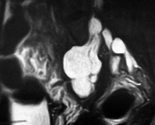
These perineural cysts are of unknown etiology and arise in sacral nerve roots, in an extradural location and communicate with the thecal sac. They are seen on pelvic ultrasound when they extend through adjacent foramina with erosion of the bone. These cysts are often multiple and bilateral. They are usually an incidental finding on asymptomatic patients, although they may cause pelvic or lower back pain.
Ultrasound Findings
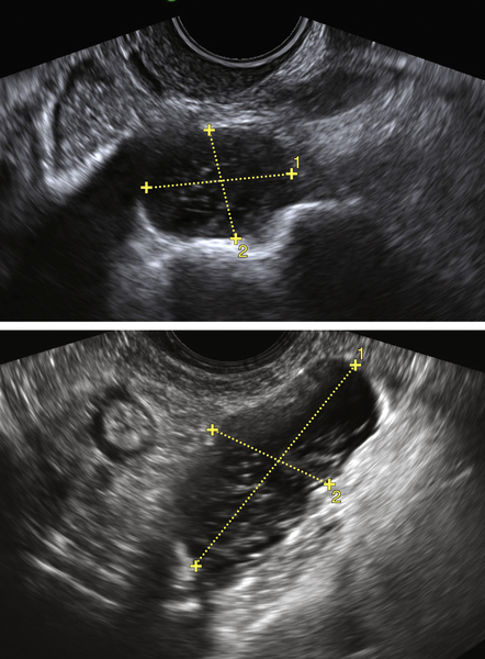
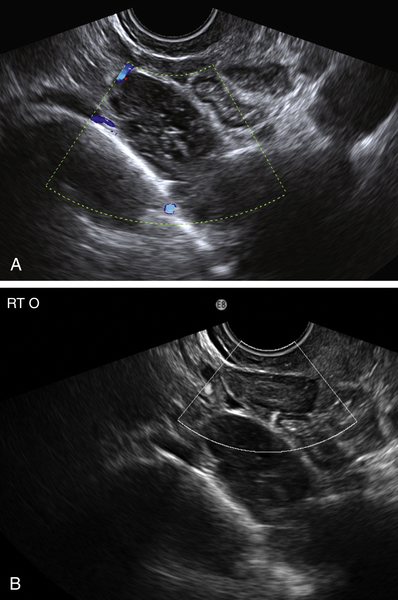
The sonographic appearance of Tarlov cysts includes cystic masses (often bilateral) in the posterior part of the pelvis, fixed to the pelvic side wall. Careful scanning will reveal that the uterus and ovaries are separate from these masses, which lie along the posterior pelvic side wall. Tarlov cysts are avascular on color Doppler.
Differential Diagnosis
It is important to visualize the ovaries separately from these cysts; otherwise, it is easy to mistake them for endometriomas, hydrosalpinges, ectopic pregnancy, lymphadenopathy (lymphoma), or retroperitoneal sarcoma. These entities all have the sonographic appearance of complex cystic masses, often bilateral and sometimes solid-looking because of their internal echoes. Because Tarlov cysts are found in the posterior compartment of the pelvis, the practitioner needs to consider this diagnosis and seek out separate ovaries to arrive at the correct diagnosis.
Clinical Aspects and Recommendations
Treatment is undertaken for symptomatic patients with perineural cysts and may involve surgery with sacral laminectomy and cyst removal. Microsurgical cyst fenestration and CT-guided percutaneous cyst aspiration have also been undertaken, but the fluid tends to reaccumulate after aspiration.
Figures
Suggested Reading
H’ng M.W., Wanigasiri U.I., Ong C.L. Perineural (Tarlov) cysts mimicking adnexal masses: a report of three cases. Ultrasound Obstet Gynecol. 2009;34:230–233.
McClure M.J., Atri M., Haider M.A., Murphy J. Perineural cysts presenting as complex adnexal cystic masses on transvaginal sonography. AJR Am J Roentgenol. 2001;177:1313–1318.
Mummaneni P.V., Pitts L.H., McCormack B.M., Corroo J.M., Weinstein P.R. Microsurgical treatment of symptomatic sacral Tarlov cysts. Neurosurgery. 2000;47:74–78.
Raza S., Klapholz H., Benacerraf B.R. Tarlov cysts: a cause of bilateral adnexal masses on pelvic sonography. J Ultrasound Med. 1994;13:803–805.


