Cystic Fibrosis

Published on 23/05/2015 by admin

Filed under Pulmolory and Respiratory

Last modified 22/04/2025

Print this page

rate 1 star rate 2 star rate 3 star rate 4 star rate 5 star
Your rating: none, Average: 0 (0 votes)

This article have been viewed 3630 times

Chapter 44 Cystic Fibrosis

Cystic fibrosis (CF) is a common, fatal, autosomal recessive disorder. Its frequency varies among populations, with approximately 1 in 3300 live births in Caucasians, 1 in 15,000 in African Americans, and 1 in 32,000 in Asians. Although reports of CF exist from medieval times, it was first described and recognized as a genetic disease by Anderson in 1938. Although the increase in sweat chloride and sodium concentrations was observed by Saint Agnese in the 1950s, it was not until 1983 that Paul Quinton described the defective chloride transport in sweat glands and respiratory epithelium as the underlying abnormality. The discovery of the causative, mutated gene encoding a defective chloride channel in epithelial cells in 1989 elucidated the pathophysiology of CF and opened up new avenues of treatment. Despite these major advances, however, it is still unclear how mutations in the cystic fibrosis transmembrane regulator (CFTR) gene precisely cause the multifaceted manifestations of CF disease.

Genetics

Cystic fibrosis is caused by mutations in a gene on chromosome 7 encoding the protein subsequently termed the CFTR gene. More than 1800 mutations have been reported to the Cystic Fibrosis Genetic Analysis Consortium. Most of these mutations are rare, and only four mutations occur in a frequency of more than 1%. CFTR mutations can be grouped into five classes: CFTR is not synthesized (I), is inadequately processed (II), is not regulated (III), shows abnormal conductance (IV), or has partially defective production or processing (V). Class I, II, and III mutations are more common and associated with pancreatic insufficiency, whereas patients with the less common class IV and V mutations often are “pancreatic sufficient” (Figure 44-1).

The most common mutation worldwide, found in approximately 66% of patients with CF, is a class II mutation caused by a deletion of phenylalanine in position 508 (F508del) of CFTR. F508del CFTR is misfolded and trapped in the endoplasmic reticulum (ER) and subsequently proteolytically degraded. However, small amounts of F508del CFTR reach the plasma membrane of epithelial cells and have some functional activity. These findings suggest that F508del CFTR rescue from ER degradation may be a potential therapeutic intervention.

The CFTR gene belongs to a family of transmembrane proteins called adenosine triphosphate (ATP)–binding cassette (ABC) transporters and functions as a chloride (Cl) channel in apical membranes. However, CFTR possesses other functions in addition to being a chloride channel. CFTR has been described as a regulator of other membrane channels, including the epithelial sodium channel (eNaC) and the outwardly rectifying chloride channel (ORCC). CFTR also transports or regulates bicarbonate (HCO3) transport through epithelial cell membranes and may act as a transporter for other proteins, such as glutathione.

A relationship exists between CFTR genotype and clinical phenotype in CF. Patients who carry two “severe” mutations (classes I, II, and III) that cause loss of function in CFTR have classic CF, characterized by pancreatic insufficiency, early age of diagnosis, and elevated sweat chloride. In contrast, patients who have at least one “mild” mutation with partial function in CFTR are typically diagnosed at an older age, have sweat chloride values closer to normal, and are pancreatic sufficient.

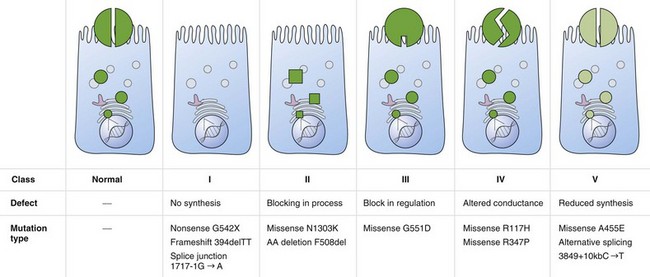
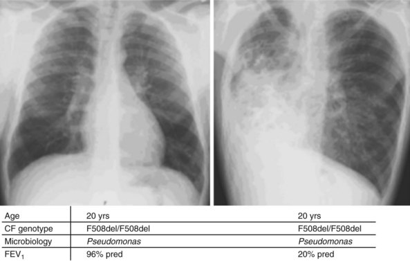
Whereas classes IV and V CFTR mutations are linked with pancreatic sufficiency, attempts to link specific mutations to the severity of lung disease have shown large phenotypic variability. This is best documented for patients homozygous for the F508del mutation who exhibit a wide spectrum in lung disease severity. This wide phenotypic variation suggests that environmental factors and genes other than CFTR influence the development, progression, and disease severity of CF (Figure 44-2).

Pathophysiology

Although there is ongoing debate on how CFTR mutations cause disease, some of the fundamental questions have been clarified in recent years. CFTR is expressed in higher quantities in tissues clinically affected by CF, such as sinuses, lungs, pancreas, liver, gastrointestinal (GI) tract, and reproductive tract, although low levels also occur elsewhere. Because lung disease is the most pertinent clinical feature of CF, the focus here is on its pathophysiology in the respiratory tract.

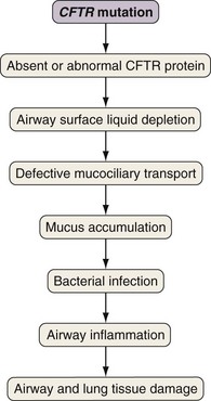
Airway epithelial cells secrete chloride and absorb sodium chloride (NaCl), the balance of which is regulated through apical channels, including CFTR (Figure 44-3). Ion secretion and absorption affect water transport, and a balance between secretion and absorption is thought to be important to maintain an adequate layer of airway surface liquid (ASL). The ASL supports the thin mucous layer on top of epithelial cells, which is constantly transported out of the lungs through ciliary movement. Lack or dysfunction of CFTR leads to reduced chloride secretion and NaCl hyperabsorption with depletion of ASL. In the absence of adequate ASL, respiratory cilia collapse, leading to breakdown of mucociliary transport. Mucus accumulates in the lower airways, and inhaled bacteria are trapped in this viscous mucous layer on top of respiratory epithelial cells.

image

Figure 44-3 Restoring airway surface liquid in cystic fibrosis.

(From Ratjen F: Restoring airway surface liquid in cystic fibrosis, N Engl J Med 354:291–293, 2006.)

The spectrum of bacteria that are relevant for CF lung disease is relatively limited. Overall, Pseudomonas aeruginosa is the most common isolate, followed by Staphylococcus aureus and Haemophilus influenzae. Later in the course of disease, multiresistant organisms such as Stenotrophomonas maltophilia, Achromobacter (Alcaligenes) xylosoxidans, and Burkholderia cepacia complex may be isolated. As in other chronic pulmonary diseases, nontuberculous mycobacteria (usually Mycobacterium avium-intracellulare or M. abscessus) may be isolated. It is challenging to prove whether these organisms are causing ongoing disease requiring treatment, or if they are colonizing only the damaged lung. For a more detailed discussion of nontuberculous mycobacteria (NTM) infections, see Chapter 31.

Stenotrophomonas maltophilia, Alcaligenes xylosoxidans, and Burkholderia cepacia complex are isolated in less than 10% of patients with CF. B. cepacia complex is an unusual organism that is found in the environment (soil and water) and causes chronic infection in only CF and chronic granulomatous disease. It is inherently multiresistant and difficult to treat and in CF is associated with a significantly worse prognosis. There is evidence for person-to-person spread in patients with CF. Approximately 15% to 20% of patients with CF who are infected with B. cepacia complex will have rapidly progressive deterioration, so-called cepacia syndrome, with necrotizing pneumonia, greatly elevated white blood cell (WBC) counts, bacteremia, and almost 100% mortality.

The mucus in CF lacks oxygen, leading to anaerobic growth conditions for bacteria. Anaerobic bacteria exist in high numbers in CF airways, but their clinical significance is uncertain. The anaerobic growth conditions trigger a switch of S. aureus and P. aeruginosa from nonmucoid to mucoid cell types, the predominant phenotype in CF lungs. These mucoid strains form biofilms in CF airways that are resistant to killing by the host defense system, resulting in chronic infection. Inflammatory products (e.g., elastase) released by neutrophils stimulate mucus secretion, perpetuating the cycle of mucus retention, infection, and inflammation.

Evidence indicates that inflammation is dysregulated in CF airways. Neutrophilic airway inflammation has been detected in infants with CF in the first months of life, as well as in CF fetal lung tissue. Whether or not inflammation is directly related to the CFTR defect is still disputed. However, an exaggerated, sustained, and prolonged inflammatory response to bacterial and viral pathogens is an accepted feature of CF lung disease. The persistent endobronchial inflammation is deleterious for the course of lung disease (Figure 44-4).

Exocrine pancreatic insufficiency is present in approximately 85% to 90% of patients with CF, generally in those patients who carry two copies of the class I, II, or III CFTR mutations. The exocrine pancreas has great functional reserve, and 98% to 99% of its function must be lost before malabsorption will occur. Patients who are pancreatic sufficient do not have normal pancreatic exocrine function but have sufficient function to prevent fat malabsorption. Pancreatic disease begins in utero and is thought to result from decreased volume of pancreatic secretions with decreased concentrations of HCO3. Without sufficient fluid and HCO3, digestive proenzymes are retained within small pancreatic ducts and are prematurely activated, ultimately leading to tissue destruction, fibrosis, and fatty replacement. The resulting malabsorption contributes to the failure to meet the increased energy demands because of the hypermetabolic state associated with endobronchial infection. Lung infections may lead to anorexia and vomiting, promoting malnutrition. These factors may exacerbate lung infection, leading to a vicious cycle of malnutrition and infection.

Clinical Features

Typical signs and symptoms for CF are listed in Box 44-1. Symptoms of CF may vary, with monosymptomatic cases often diagnosed late. It is therefore important to be aware of the spectrum of symptoms that may arise and to initiate adequate diagnostic steps.

Although the classic presentation of CF is the combination of chronic productive cough, steatorrhea, and failure to thrive, 10% to 15% of patients do not have pancreatic insufficiency clinically. Because the lungs of patients with CF are normal at birth, pulmonary symptoms may not be obvious. Between 10% and 15% of newborn infants with CF may fail to pass meconium, leading to meconium ileus, which is linked to pancreatic insufficiency but not directly associated with more severe clinical disease.

Less common presentations of CF, such as prolonged jaundice in the newborn and rectal prolapse in infants and young children, should trigger diagnostic tests. Occasionally, an infant may have severe malnutrition with anemia, hypoalbuminemia, and edema in the first 4 months of life.

Infertility can be a presenting symptom in adult patients with CF with limited pulmonary symptoms; 98% of males with CF are infertile, with azoospermia secondary to atretic or absent vas deferens. Spermatogenesis and sexual potency are normal. Female reproductive function is normal, although a lower rate of fertility has been postulated because of dehydrated cervical mucus.

Initially, pulmonary symptoms of cough will occur only at times of exacerbations, but eventually there is progression to a chronic daily cough productive of sputum. The sputum is initially white, but as infection continues, the mucus becomes thicker and purulent. Minor hemoptysis often occurs at exacerbation. Some patients have an “asthmatic” component to their disease, with wheezing, chest tightness, paroxysmal dry cough, and a degree of reversibility in airflow obstruction with bronchodilators. Over time, as pulmonary function declines, there is increasing dyspnea. Hypoxemia is not usually seen until forced expiratory volume in 1 second (FEV1) is less than 35% of predicted value, and hypercarbia usually occurs when the FEV1 is less than 25% or 30% predicted. Cor pulmonale occurs late in the illness.

Pansinusitis is found on sinus radiographs in most patients, although not all will have symptoms of recurrent sinusitis, headache, and postnasal drip. Nasal polyps are seen in approximately 20% of patients and tend to recur even after surgical removal.

Diagnostic Approach

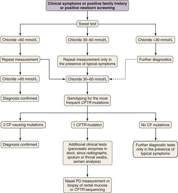
The diagnosis of cystic fibrosis is established by clinical manifestations (see Box 44-1), a history of CF in a sibling, or a positive newborn screening result, in conjunction with laboratory evidence of CFTR dysfunction. CFTR dysfunction is documented by elevated sweat chloride or characteristic abnormalities in nasal potential difference or by CF-causing mutations in the CFTR gene.

A diagnostic algorithm is presented in Figure 44-5. Abnormal ion transport is reflected in high sweat NaCl levels, and measurement of chloride concentration in sweat after iontophoresis of pilocarpine is used for diagnosis. Sweat testing must be done using standardized methods, by qualified staff in an experienced laboratory. A sweat chloride concentration greater than 60 mmol/L on repeated analysis is diagnostic for CF; 30 to 60 mmol/L is considered a “borderline” result but may be seen in patients with CF.

Diagnosis can be confirmed by genotyping of the most common CFTR mutations, which vary by ethnic origin of the population tested. More than 1800 mutations in CFTR have been reported to the CFTR database. Most commercial screening panels test for less than 50 mutations and will identify 85% to 90% of CF alleles. Although CFTR mutation testing has had no clinical implications in the past, this may change with the introduction of mutation-specific therapy (see later discussion).

The diagnosis of CF requires the presence of two CF-causing mutations. To be considered CF-causing, the mutation must (1) cause a change in the amino acid sequence that severely affects CFTR synthesis or function, (2) introduce a premature termination signal (insertion, deletion, or nonsense mutations), (3) alter the “invariant” nucleotides of intron splice sites (first or last two nucleotides), or (4) cause a novel amino acid sequence that does not occur in the normal CFTR genes from at least 100 carriers of CF mutations from the patient’s ethnic group. Of the more than 1800 CFTR mutations reported, only approximately 25 are considered disease-causing to date.

If CFTR genotyping or sweat test is not diagnostic, a second test of CFTR function such as nasal potential difference (NPD) measurement can be performed. The transport of Na+ and Cl ions across the nasal mucosa creates a transepithelial electrical potential difference. Changes in NPD in response to stimulation or inhibition of ion channels by nasal perfusion can be measured, and a typical normal or CF response exists. The NPD test is technically difficult, requiring a skilled operator, and therefore is not available in all CF centers. Standard operating procedures and reference values have recently been determined. Other techniques to examine CFTR function include analysis of rectal mucosal biopsies in an Ussing chamber.

Clinical tests not directly assessing the CFTR defect can also aid in the diagnostic process. Most patients with CF are pancreatic insufficient, and a decreased concentration of chymotrypsin or pancreas-specific elastase in feces or 72-hour stool collection with fecal fat analysis can confirm this. Most patients with CF have total opacification of paranasal sinuses, and sinus radiography may be helpful. Bacterial pathogens typical for CF (e.g., mucoid Pseudomonas, S. aureus) can be detected in sputum or throat swabs and suggest a CF diagnosis. Obstructive azoospermia is found in 98% of men with CF and is a result of congenital bilateral absence of the vas deferens (CBAVD). The finding of azoospermia, or lack of vas deferens on careful urologic examination or transrectal ultrasound, suggests CF.

In the past, about 50% of patients with CF in North America were diagnosed by age 6 months and 90% by 8 years. Neonatal screening has been proposed as early diagnosis, and therefore earlier initiation of treatment may improve outcome. A randomized screening program in Wisconsin state found that weight gain and early growth were better in patients diagnosed by neonatal screening. Because good nutrition is linked to a better prognosis, these data would favor the introduction of population-wide neonatal screening. Neonatal screening programs have now been introduced in many countries, most often based on a two-step approach, with immunoreactive trypsinogen (IRT) in dried blood spots and confirmation by DNA analysis in positive cases. Blood trypsinogen is elevated in pancreatic-insufficient patients in the first weeks of life, but the rate of false-positive results of a single IRT test is high, which can be reduced substantially by inclusions of a second step, including genetic testing for the most common CFTR mutations.

Diagnostic Challenges

In 5% to 10% of patients, the diagnosis of CF is not made until adulthood. Increased awareness continues to result in more adults being diagnosed with CF. Most will not have typical features of CF but rather single-organ disease (CBAVD, recurrent acute pancreatitis, or bronchiectasis) or symptoms that develop later in life. Most patients diagnosed as adults are pancreatic sufficient, have “borderline” sweat chloride tests, and have mutations in the CFTR gene that are not considered CF-causing. Commercial genetic screening panels may have diagnostic limitations for patients seen in adulthood by including the more common CF-causing mutations usually seen in childhood diagnosis. Adults are more likely to have less common CFTR mutations that are not part of the panel. Complete sequencing of the CFTR gene has now become feasible and may offer additional information to assist in the diagnosis of CF, especially for patients with equivocal sweat test results. NPD measurement may be especially helpful in establishing a diagnosis of CF in these patients. Interpretation of these results should be done by an experienced CF physician, who is aware of the limitations of these tests.

Obstructive azoospermia is highly related to mutations in CFTR, and the finding of CBAVD should trigger genetic testing for CF, especially because assisted reproductive techniques allow these men to father children. Up to 80% of men with CBAVD may have one or two CFTR mutations but no other phenotypic features of CF. A diagnosis of CF should not be made in these cases unless diagnostic criteria are met (sweat test >60 mmol/L or 2 CF-causing mutations). The long-term prognosis for these men is still not certain, including whether typical CF lung disease may develop over time, and thus they should receive appropriate clinical follow-up. The relationship between CFTR mutations and recurrent acute pancreatitis, bronchiectasis, or other sinopulmonary disease is not as strong as that seen with CBAVD. However, a higher-than-expected prevalence of CFTR mutations is found in these populations.

Despite all these tests, a small number of patients remain in whom a definite diagnosis cannot be made. Typical CF bronchiectasis may be seen in patients who have CFTR mutations that are not considered disease-causing and who have a normal or borderline sweat chloride test. Thus, it seems that CF lung disease may occur even with lesser degrees of dysfunction of CFTR. The important message is that presence of CFTR mutations may put patients at risk for significant CF disease developing later in life, and close follow-up is advised.

This dilemma is further complicated by lack of a common and standardized terminology for patients who have evidence of CFTR dysfunction without a definite diagnosis of CF. These patients are being referred to as atypical CF, mild CF, nonclassic CF, or CFTRopathy, or if CFTR mutations exist in the absence of lung or GI disease, pre-CF. The current preferred term for patients with a CF clinical phenotype and evidence of CFTR dysfunction but do not meet diagnostic criteria for CF is CFTR-related disorder.

Clinical Course of Lung Disease and Principles of Therapy

The lungs of newborns with CF are normal at birth. Pathologic studies show that the first abnormalities are usually detected in the small airways reflected by mucous plugs and dilation distal to the obstructed airways. These early abnormalities are often focal and do not necessarily produce clinically apparent symptoms. The disconnect between pathologic abnormalities and pulmonary symptoms is now recognized and has important implications for treatment. Studies that use bronchoalveolar lavage fluid show that ongoing airway inflammation is present in patients with mild disease and can be observed early in infancy. The lack of impressive symptoms does not mean that these patients do not have ongoing progressive disease requiring continuous treatment.

Although the traditional approach was to wait for symptoms, a more aggressive strategy is used today. This aggressive approach translates into continuous lifelong treatment, which is time-consuming and labor-intensive for patients and their families. Although the burden for individuals is considerable, it has changed the natural history of disease progression in patients with CF. The classic course of CF was characterized by chronic productive cough and a gradual, but steady, decline in pulmonary function Now most patients with CF maintain their pulmonary function over years, reflected in an annual rate of decline in FEV1 of less than 2% predicted per year, and many do not regularly expectorate sputum until adulthood. However, it is important to be vigilant about periods of deterioration that present as episodes of pulmonary exacerbations and, unless treated rapidly and aggressively, may lead to an irreversible loss in pulmonary function.

Pulmonary exacerbations in patients with CF are often triggered by viral infections but require prompt treatment with antibiotics directed against pathogens present in respiratory cultures. It is important to recognize the signs of a pulmonary exacerbation early to avoid permanent damage to the lung (Box 44-2). In general, any increase in symptoms lasting more than a few days, associated with a decline in FEV1, requires antibiotic therapy. Because most CF patients are chronically infected with bacteria, suppression rather than eradication of organisms is the primary goal of therapy. Duration of therapy is not guided by changes in sputum bacteriology but rather by improvement in symptoms and pulmonary function.

Because many patients have normal lung function and the annual rate of decline in pulmonary function is minimal, it has become difficult to assess lung disease by lung function alone. Despite this, FEV1 remains the best predictor of outcome in patients with CF and is therefore used to follow the course of lung disease and to guide treatment. Chest radiographs are performed annually in most centers but have limited sensitivity to detect early abnormalities. High-resolution computed tomography can detect abnormalities not detected by conventional radiography, but CT is associated with a higher cumulative radiation dose and thus not considered part of routine care at present. There is considerable interest in developing more sensitive outcome parameters assessing function, structure, infection, and inflammation to help guide treatment, but currently these tests have a limited role outside of research studies.

Treating bacterial infections and avoiding lung function decline are key to management of CF patients, with regular assessment of clinical status, lung function, and sputum microbiology. Quarterly clinic visits are considered standard in CF care, but some patients require more frequent follow-up.

Treatment

Gene Replacement Therapy and Pharmacotherapy

Cystic fibrosis is caused by deficient or absent CFTR, and thus gene replacement therapy is a form of curative treatment. Trials so far have targeted the respiratory tract directly. A number of vector systems have been tested in human trials, but adenoviruses and cationic lipids are the two most common vectors. Even though some transient effect on CFTR expression and function has been achieved, no study could demonstrate a long-lasting effect. Viral vectors seem to be more efficient than cationic lipids but have the disadvantage of being immunogenic. Although attempts are ongoing to overcome these shortcomings, gene therapy is currently not a therapeutic option for patients with CF.

Another approach to treat the underlying defect is CFTR pharmacotherapy aiming to improve trafficking, expression, or function of CFTR. For patients carrying class I stop mutations, which lead to premature termination codons in the messenger RNA and therefore truncated protein production, treatment with aminoglycosides or its derivative ataluren has been shown to increase CFTR expression. Uncontrolled studies also show improved chloride channel function in the nasal epithelium as well as positive trends in lung function. A larger placebo-controlled trial now underway will further clarify clinical benefits of this therapy.

Because most patients do not carry stop mutations, this approach addresses only a small fraction of the CF population. A potentiator of CFTR function, VX770, has been tested in patients with the G551D mutation, in which CFTR is present on the cell surface but channel function is reduced. Recent studies demonstrate that treatment not only improved lung function, but also reduced sweat chloride concentrations below the threshold clearly diagnostic for CF. A subsequent longer study confirmed these results, and VX770 has become the first CFTR modulator to be licensed for clinical use.

For the most common mutation, F508del, misfolded CFTR is degraded in the ER before reaching the cell membrane. Because this misfolded CFTR does have Cl-conducting function, compounds that affect intracellular trafficking, called chaperones, may provide clinical benefit. Some studies have provided proof of concept, and one compound derived from high-throughput screening programs, VX809, is currently tested in clinical trials. Safety has been confirmed in early trials. Because the CFTR potentiator VX770 greatly enhanced the effect of CFTR corrector therapy in vitro, combination therapy with VX809 is also being tested to enhance therapeutic efficacy.

An alternative to CFTR pharmacotherapy is to activate other Cl channels present in the apical surface of epithelial cells or to inhibit Na+ hyperabsorption. This concept seems useful because CF mice lacking CFTR do not develop lung disease, which may be caused by better function of alternative Cl channels. Whether activation of alternative Cl channels or inhibition of Na+ hyperabsorption is a valid treatment option is currently unclear. Denufosol, an activator of alternative chloride secretion, has undergone a full development program and showed promise in improving lung function in both Phase II studies and a 6-month placebo-controlled Phase III trial. However, the second Phase III trial failed to confirm these findings.

Symptomatic Therapy

In the absence of a proven curative regimen, symptomatic treatment is still the mainstay of CF therapy. Most of the treatment approaches are directed at interrupting the cycle of mucus retention, infection, and inflammation. Early initiation of therapy is important to avoid permanent damage to the lung.

Airway Clearance

Chest physiotherapy remains the mainstay of airway clearance and is recommended for all patients. Short-term benefits have been demonstrated for many techniques, but long-term efficacy data are limited. The most common techniques are manual percussion, positive expiratory pressure (PEP) mask therapy, and autogenic drainage. In addition, techniques that provide vibration to the airways actively or passively are being used. Debate is ongoing as to which technique provides the best efficacy for patients with CF. Physical activity and exercise are considered important adjuncts to physiotherapy, because impairments in exercise tolerance have been linked to poorer prognosis in CF patients.

Airway secretions in CF patients are highly viscous, indicating the use of drugs that reduce the viscoelasticity of sputum. Classic mucolytics such as N-acetylcysteine have little effect on lung disease in CF, although they are being revisited because of their potential benefit as antioxidants. Their ineffectiveness as a mucolytic may be because CF mucus contains little mucin and is mainly composed of pus. Recombinant human deoxyribonuclease (rhDNase) administered by inhalation reduced sputum viscosity, improved pulmonary function, and reduced the number of pulmonary exacerbations in patients with moderate and with mild lung disease. Data also suggest that rhDNase reduces inflammation in the airways. Hypertonic saline has been assessed as another potential drug to improve airway clearance, and recent studies suggest that it may also increase airway surface liquid. The effect on lung function seems to be smaller than that of rhDNase, but these therapies have different mechanisms of action and thus cannot be viewed as virtually exclusive. Hypertonic saline also reduces the frequency of pulmonary exacerbations, an important outcome because these are associated with decreased lung function.

Treatment of Airway Infection

Aggressive treatment of airway infection is a main reason for the increased life expectancy of patients with CF achieved over recent decades. As mentioned, bacterial pathogens in patients with CF are usually limited to a relatively small spectrum, with S. aureus or H. influenzae the most prominent in younger patients and P. aeruginosa in older patients. Most patients go through phases of clinical stability with intermittent pulmonary exacerbations. A set of criteria is used to diagnose pulmonary exacerbations (see Box 44-2), but the threshold for initiating targeted antibiotic therapy should be rather low. Genotyping has shown that the organisms present at exacerbation are the same as when the patient is clinically stable, but with higher bacterial density. Thus, choice of antimicrobial therapy on the basis of the most recent sputum cultures is indicated.

Pulmonary exacerbations are usually treated with intravenous (IV) antibiotic therapy, although oral therapy is used for exacerbations associated with minimal or no drop in lung function. Antistaphylococcal antibiotics are usually administered for 2 to 4 weeks. Patients with CF have differences in drug clearance and require dosages approximately 50% higher than individuals without CF. For patients with Pseudomonas infection, combination therapy with a semisynthetic penicillin (e.g., piperacillin), a third-generation cephalosporin (e.g., ceftazidime), or a carbapenem (imipenem or meropenem) with an aminoglycoside (most frequently tobramycin) is administered for 2 to 3 weeks. Oral ciprofloxacin is used for less severe exacerbations.

Treatment of the less common gram-negative organisms such as B. cepacia complex, A. xylosoxidans, and S. maltophilia, can be challenging. These organisms are inherently multiresistant. Although infection with B. cepacia complex is clearly associated with a worse prognosis, the relevance of the other gram-negative bacteria is less clear, and currently it is not established whether they are responsible for causing disease. For patients chronically infected with B. cepacia complex, trimethoprim-sulfamethoxazole (TMP-SMX) or doxycycline is effective for minor exacerbations. For more severe infections, the best in vitro antibiotics consist of meropenem, and high-dose inhaled tobramycin in combination with either ceftazidime, chloramphenicol, or TMP-SMX. Treatment of exacerbations with B. cepacia complex may require prolonged antibiotic therapy (weeks to months) before a clinical response is seen. Use of pulmonary function, WBC count, and markers of inflammation (CRP or ESR) may be helpful to guide therapy. S. maltophilia can be treated with TMP-SMX or doxycycline. Because S. maltophilia isolates can develop resistance during treatment, TMP-SMX is often combined with a second antibiotic, such as ticarcillin-clavulanate or levofloxacin. A. xylosoxidans can be challenging to treat, but options include imipenem and piperacillin. Inhaled colistin may also be effective, because in vitro studies found that high concentrations of colistin inhibit most strains of A. xylosoxidans.

Many CF physicians try to eradicate bacteria from CF airways with courses of oral antibiotics even in the absence of symptoms. Prophylactic antistaphylococcal therapy with flucloxacillin initiated at diagnosis is also used in some centers. Although one small study has reported a lower rate of cough and hospital admissions during the first 2 years of life, continuous antistaphylococcal therapy was associated with a higher rate of P. aeruginosa acquisition in two other studies, in which mainly cephalosporins were used. P. aeruginosa infection increases pulmonary inflammation and has a negative effect on lung function when this pathogen persists. At present, there is insufficient evidence to support the use of prophylactic antistaphylococcal therapy in patients with CF.

Overall, Pseudomonas aeruginosa is the major pathogen in CF lung disease. Its prevalence increases with age, and most adult patients are chronically infected with this organism. After an initial transient colonization period with nonmucoid strains, untreated patients generally become chronically infected with mucoid strains of P. aeruginosa. Antibiotic therapy usually fails to eradicate mucoid P. aeruginosa from the airways. High bacterial counts, low metabolic rate of pathogens in biofilms, poor penetration of antibiotics into airway secretions, and anaerobic conditions in sputum are considered responsible for this finding. Chronic infection with mucoid strains has a negative impact on the subsequent course of lung disease. Although eradication is virtually impossible in chronic infection, treatment can be effective in the early phase of P. aeruginosa infection; a major improvement in patients with CF is early antibiotic therapy. Both inhaled antibiotic therapy with tobramycin alone and combined inhaled antibiotics and oral ciprofloxacin have been used, but more recent evidence suggests that adding ciprofloxacin does not increase therapeutic success. Although the optimal treatment regimen for early P. aeruginosa infection has yet to be determined, inhaled tobramycin successfully reduces the incidence of chronic airway infection with P. aeruginosa in patients with CF (Table 44-1).

Table 44-1 Options for Oral and Inhaled Antibiotic Therapy

Antibiotic: Choose One Pediatric Dose Adult Dose
Staphylococcus aureus
Dicloxacillin 6.25-12.5 mg/kg four times daily 250-500 mg four times daily
Cephalexin 12.5-25 mg/kg four times daily 500 mg four times daily
Amoxicillin/clavulanate 12.5-22.5 mg/kg amoxicillin* 400-875 mg amoxicillin*
Haemophilus influenzae
Amoxicillin 25-50 mg/kg twice daily 500-875 mg twice daily
Amoxicillin/clavulanate 12.5-22.5 mg/kg amoxicillin* 400-875 mg of amoxicillin*
Cefuroxime axetil 15-20 mg/kg twice daily 250-500 mg twice daily
Pseudomonas aeruginosa
Ciprofloxacin 10-15 mg/kg twice daily 750 mg twice daily
Tobramycin 300 mg by nebulizer, twice daily 300 mg by nebulizer, twice daily
Colistin 150 mg by nebulizer, twice daily 150 mg by nebulizer, twice daily

* Component twice daily.

By inhalation.

Modified from Gibson RL, Burns JL, Ramsey BW: Am J Respir Crit Care Med 168:918–951, 2003.

To avoid adverse effects and to obtain high drug concentrations in airways, inhaled antibiotic therapy is the treatment of choice for maintenance therapy in patients with P. aeruginosa infection. The best evidence currently available is for inhaled tobramycin, which has been shown to improve lung function and reduce pulmonary exacerbations in chronically infected patients. In addition, colistin is being used “off label” for inhalation. Although colistin has the advantage of low prevalence of resistant strains, its short-term efficacy is inferior to inhaled tobramycin. Inhaled aztreonam has been recently licensed as an alternative option to tobramycin based on two controlled clinical trials demonstrating its efficacy. A comparison to inhaled tobramycin also found similar efficacy with aztreonam and suggested superior lung function benefits during therapy; however, most of the patients were previously treated with inhaled tobramycin, which may favor the response to aztreonam. Both tobramycin and aztreonam have been studied in cycles of 28 days on and 28 days off, so many clinicians have also started to use both treatments in alternate months to avoid deterioration during the off periods.

Some centers treat chronically infected CF patients with routine IV antibiotic therapy every 3 months, regardless of respiratory symptoms, but this approach has not yet been supported by sufficient evidence.

In addition to inhaled antibiotics, azithromycin can improve pulmonary function and reduce pulmonary exacerbations in patients with P. aeruginosa –positive disease and more recently has been shown to reduce exacerbations in P. aeruginosa–negative patients as well. Although macrolides have no efficacy against P. aeruginosa when tested in routine cultures, some evidence suggests macrolides may affect P. aeruginosa growing in biofilms. Whether this explains their efficacy or whether this is caused by antiinflammatory properties of macrolides is still unclear (Table 44-1).

Patient-to-patient transmission of bacterial pathogens does occur. Thus, separation regimens have been implemented to prevent cross-infection in patients with CF. This is particularly important with B. cepacia complex because person-to-person transmission has been proved, and chronic infection is associated with a worse clinical outcome. Vaccines are being developed against P. aeruginosa, but their efficacy is currently unproven.

Complications

Pulmonary Complications

Hemoptysis

Chronic pulmonary infection leads to enlargement of the bronchial artery circulation supplying the lung. Hemoptysis can occur even in patients with mild lung disease and usually indicates infection. Vitamin K deficiency caused by pancreatic insufficiency can contribute to the problem. Massive hemoptysis (>250 mL of blood in 24 hours) is less common but occasionally can be life-threatening (see Chapter 20). Hemoptysis is usually treated with antibiotic therapy. Vitamin K and tranexamic acid may also be used. Ongoing massive hemoptysis requires bronchial artery embolization. Angiography is used to locate the abnormal bronchial artery vessels. These vessels are aberrant, and arteries arising from one side may supply contralateral segments of the lung. Because location of the bleeding does not always correlate with origin of abnormal bronchial arteries, bronchoscopy to locate source of bleeding is not helpful. However, bronchoscopy may be necessary to manage the airway in life-threatening hemoptysis. Embolization should be performed by experienced interventional radiologists because of potential complications of bronchial artery embolization, including infarction of the esophagus, lung parenchyma, or chest wall (causing dysphagia or severe chest pain), and transverse myelitis caused by accidental embolization of the spinal arteries.

Gastrointestinal Complications

Distal Intestinal Obstruction Syndrome

Undigested fat, mucus from the GI tract, swallowed sputum, and reduced water in the small bowel result in thick, sticky bowel contents. Partial bowel obstruction, usually at the ileocecal junction, can cause recurrent right lower quadrant (RLQ) pain and occasionally altered bowel habit. This condition occurs in 15% to 20% of patients and is called distal intestinal obstruction syndrome (DIOS). The diagnosis is made clinically from the history and palpation of a tender mass in the RLQ along with evidence of fecal collection in the RLQ on abdominal imaging. DIOS rarely causes complete bowel obstruction, elevated WBC count, or fever; if these features are present, other causes of abdominal pain should be considered, such as intussusception, appendicitis or appendiceal abscess, and Clostridium difficile colitis. Treatment requires large volumes of intestinal lavage solution (GoLytely, Peglyte) to clear the bowel. Occasionally, enemas with water-soluble osmotic agents such as Gastrografin or Hypaque (diatrizoate sodium) are needed. Prevention of recurrent DIOS should focus on adequate pancreatic enzyme replacement and use of mineral oil or polyethylene glycol.

Intussusception can occur in 1% to 2% of children and young adults with CF and may mimic DIOS. The location of the intussusception is usually ileo-ileo but may be ileo-colic. Intussusception may be recurrent and asymptomatic but can cause small bowel obstruction. It also may be associated with intermittent severe colicky abdominal pain, a palpable mass, and vomiting.

Cystic Fibrosis–Related Bone Disease

Adults with CF have evidence of decreased bone density, including osteopenia and osteoporosis, as measured by bone dual-energy x-ray absorptiometry (DEXA). Prevalence of low bone density in adults with CF ranges from 40% to 70%, and up to 50% of adults with severe lung disease who are awaiting transplantation will have osteoporosis. The etiology of the decreased bone density is multifactorial, and both failure of normal bone formation and excessive bone loss occur. Malnutrition, vitamin D and K deficiency, hypogonadism, increased bone loss because of elevated inflammatory cytokines from chronic pulmonary infection, and use of corticosteroids all may play a role in the development of low bone density. It is not completely clear whether DEXA scans predict fracture risk in patients with CF. A consensus report suggests treatment with calcium and vitamin D supplementation, treatment of hypogonadism if present, promotion of exercise, treatment of pulmonary infection, and use of bisphosphonates in patients with osteoporosis, in those receiving corticosteroids, and in those on the transplant list.

Hypertrophic pulmonary osteoarthropathy (HPOA) is a chronic proliferative periostitis associated with digital clubbing. It can cause severe bony pain and swelling, especially in knees, ankles, and wrists. It frequently flares at times of pulmonary exacerbations, improving with treatment of the lung infection. Radiography confirms the typical periosteal new bone formation at the distal ends of long bones.

Episodic arthropathy occurs in 5% to 10% of CF patients and presents as transient episodes of acute swelling and joint pain involving single or multiple, large or small joints. These episodes do not necessarily correlate with pulmonary exacerbations. This is a nonerosive arthropathy and responds to nonsteroidal antiinflammatory drugs or corticosteroids.

About 1% to 2% of patients with CF develop a cutaneous vasculitis, which on biopsy is a leukocytoclastic vasculitis. Systemic involvement is rare. It is usually seen on the lower extremities, and the rash consists of usually painless purpura. This typically resolves spontaneously, and specific treatment is not required.