5.1 Cyanotic heart disease and tetralogy of Fallot spells
Introduction
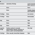
Cyanosis is a bluish discolouration of skin and mucous membranes due to excessive concentration of reduced haemoglobin in the blood.1 Cyanosis is evident when deoxygenated haemoglobin in the cutaneous veins reaches approximately 5 g dL–1.2 Deoxygenated haemoglobin may occur either from arterial blood desaturation or increased oxygen extraction by peripheral tissue. Central cyanosis is produced as a result of arterial desaturation, i.e. aortic blood carrying deoxygenated haemoglobin. Isolated peripheral cyanosis may result from excessive deoxy-haemoglobin caused by extensive oxygen extraction.1 Haemoglobin level affects the presence of cyanosis. Cyanosis is detected at a higher oxygen saturation in children with polycythaemia and is more difficult to detect in children with severe anaemia. Causes of cyanosis are listed in Table 5.1.1.
| Differential diagnosis |
| Arterial oxygen desaturation (central cyanosis [pO2 <50 mmHg]) |
Cyanotic congenital heart disease
Arterial oxygen saturation should always be assessed with pulse oximetry when considering cyanosis in the newborn. The hyperoxia test may help distinguish cyanosis caused by cyanotic heart defects from other defects.2–4,8
Management
Treatment2–6,8–13


1 Anderson D.M. Dorland’s Illustrated Medical Dictionary. Philadelphia: WB Saunders; 1994.
2 Park M.K. Congestive heart failure. In Park M.Y., editor: Pediatric cardiology for practitioners, 5th ed., Philadelphia: Mosby Elsevier, 2008.
3 Abelson W.H., Garth Smith R. Residents handbook of pediatrics, 7th ed. Toronto: The Hospital for Sick Children, Toronto, Canada, BC Decker Inc; 1987.
4 Guzman M.F., Hedley Brown A., Been M., et al. Manual of cardiorespiratory critical care. Sevenoaks, Kent: Butterworth; 1989.
5 Kilham H., Isaacs D. The New Children’s Hospital Handbook. Westmead: RAHC; 1999.
6 Apitz C., Webb G.D., Redington A.N. Tetralogy of Fallot. Lancet. 2009;374:1462-1471.
7 Allen H.D., Driscoll D.J., Shaddy R.E., et al. Moss and Adams’ heart disease in infants. children and adolescents: Including the fetus and young adult, 7th ed. Philadelphia: Lippincott Williams & Wilkins; 2007.
8 Nichols D.G., Cameron D.E. Critical heart disease in infants and children, 2nd ed. Philadelphia: Mosby Elsevier; 2006.
9 Siwik E.S., Erenberg F., Zahka K.G., Goldmuntz E. Tetralogy of Fallot. In Allen H.D., Driscoll D.J., Shaddy R.E., et al, editors: 2007 Moss and Adams’ Heart Disease in Infants, Children and Adolescents: Including the Fetus and Young Adult, 7th ed., Philadelphia: Lippincott Williams & Wilkins, 2007.
10 Park M.K. Tetralogy of Fallot. In Park M.Y., editor: Pediatric cardiology for practitioners, 5th ed., Philadelphia: Mosby Elsevier, 2008.
11 van Roekens C.N., Zuckerberg A.L. Emergency management of hypercyanotic crises in tetralogy of Fallot. Ann Emerg Med. 1995;25(2):256-258.
12 Baele P.L., Rennotte M.T., Veyckemans F.A. External compression of the abdominal aorta reversing tetralogy of Fallot cyanotic crisis. Anaesthesiology. 1991;75(1):146-149.
13 Nussbaum J., Zane E.A., Thys D.M. Esmolol for the treatment of hypercyanotic spells in infants with tetralogy of Fallot. J Cardiothorac Anaesthesiol. 1989;3(2):200-202.
Allen H.D., Driscoll D.J., Shaddy R.E., et al. Moss and Adams’ heart disease in infants, children and adolescents: Including the fetus and young adult, 7th ed., Philadelphia: Lippincott Williams & Wilkins, 2007.
Apitz C., Webb G.D., Redington A.N. Tetralogy of Fallot. Lancet. 2009;374:1462-1471.
Breitbart R.E., Fyler D.C. Tetralogy of Fallot. In Keane J., Fyler D., Lock J., editors: Nadas paediatric cardiology, 2nd ed., Philadelphia: Saunders Elsevier, 2006.
Nichols D.G., Cameron D.E. Critical heart disease in infants and children, 2nd ed, Philadelphia: Mosby Elsevier, 2006.
Park M.K. Congestive heart failure. In Park M.Y., editor: Pediatric cardiology for practitioners, 5th ed., Philadelphia: Mosby Elsevier, 2008.
van Roekens C.N., Zuckerberg A.L. Emergency management of hypercyanotic crises in tetralogy of Fallot. Ann Emerg Med. 1995;25(2):256-258.