Cyanosis
Cyanosis is the abnormal blue discoloration of the skin and mucous membranes resulting from the presence of 5 g/dL or more of reduced haemoglobin in the blood. It is not synonymous with hypoxaemia, which may be present (e.g. anaemia) without cyanosis.
History
Central cyanosis
Onset
Cyanosis due to congenital heart disease causing anatomical right to left shunts may have been present from birth or the first few years of life. Immediate onset of cyanosis may be due to pulmonary emboli or cardiac failure. Acute onset of cyanosis may be precipitated by pneumonia and asthma. Patients with COPD develop cyanosis over many years. Accompanying polycythaemia may exacerbate cyanosis in these patients.
Chest pain
Cyanosis associated with pleuritic chest pain may be due to pulmonary emboli or pneumonia. Dull, aching chest tightness is experienced by patients who develop cyanosis from pulmonary oedema as a complication of myocardial infarction.
Dyspnoea
Sudden onset of dyspnoea can occur with pulmonary emboli and pulmonary oedema, while conditions such as asthma may produce a more gradual onset.
Past medical history and drug history
Any co-existing respiratory disease is significant, as cyanosis can result from any lung disease of sufficient severity. Consumption of drugs such as phenacetin and sulphonamides may precipitate methaemoglobinaemia and sulphaemoglobinaemia, respectively.
Peripheral cyanosis
General history
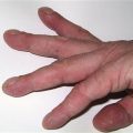
Acrocyanosis is a condition in which the hands are persistently blue and cold; it is not associated with pain. Raynaud’s phenomenon is the episodic three-colour change that occurs, with arterial vasospasm (white), cyanosis (blue) and reactive hyperaemia (red). It may be idiopathic or be associated with connective tissue diseases and drugs such as beta blockers.
Peripheral cyanosis may also result from acute arterial occlusion and is accompanied by pain and mottling of the skin. Iliofemoral deep venous thrombosis can produce a painful blue leg, termed phlegmasia cerulea dolens.
Examination
Temperature
Pneumonia and pulmonary emboli may be associated with pyrexia.
Inspection
It is often difficult to detect minor degrees of cyanosis. Central cyanosis produces a blue discoloration of the mucous membranes and digits; peripheral cyanosis produces blue discoloration only of the digits. Episodic peripheral cyanosis may be due to Raynaud’s disease and this may be associated with small areas of infarction on the fingertips. The presence of clubbing may be due to congenital cyanotic heart disease. Classically, patients with chronic bronchitis appear cyanosed with a poorly expanding barrelled chest. The JVP is elevated with congestive cardiac failure.
Respiratory examination
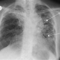
Poor chest expansion occurs with chronic bronchitis and asthma. Unilateral impairment of expansion may occur with lobar pneumonia; in addition, dullness to percussion is experienced over the area of consolidation. Localised crepitation may be auscultated with lobar pneumonia, but is more widespread with bronchopneumonia, pulmonary oedema and chronic bronchitis. Air entry is poor with chronic bronchitis and asthma. Bronchial breathing may be auscultated over an area of consolidation, and additional sounds, such as wheezing, may be heard with asthma.
General Investigations
■ Oxygen saturation
Saturation is usually below 85% with cyanosis (unless methaemoglobinaemia where saturation is normal).
■ ABGs
↓ pO2 all severe lung disease.
■ FBC
Hb ↑ chronic cyanosis. WCC ↑ pneumonia and pulmonary embolism.
■ ECG
Features of myocardial infarction (p. 62). Non-specific abnormalities with pulmonary emboli.