131
Cutaneous T-cell lymphoma
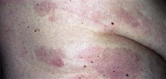
Early lesions are red with subtle scale or fine wrinkles. They persist for months with little peripheral extension and are often diagnosed as eczema.
Infiltration of the entire skin produces thickened red skin with or without scale. These features are subtle, not always clinically diagnostic, and may require a biopsy for diagnosis.
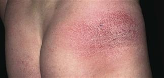
Pink to red, slightly scaly patches of early mycosis fungoides; may be present for months or years, and misdiagnosed as eczema. Biopsy can help differentiate these subtle changes of cutaneous T-cell lymphoma.
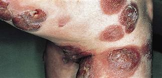
Tumors develop from pre-existing plaques or erythroderma. Tumors can vary in size; some become huge.
DESCRIPTION
Also known as mycosis fungoides, this is a distinct helper T-cell lymphoma of skin. T-lymphocytes invade the skin, lymph nodes, peripheral blood, internal organs.
HISTORY
• Evolves through several stages: early or pre-mycosis fungoides, patch, plaque, tumor stage. Stage varies from patient to patient. • Sézary syndrome is the blood or leukemic form. More common in men, African-Americans. Typically diagnosed in fifth or sixth decade.
PHYSICAL FINDINGS
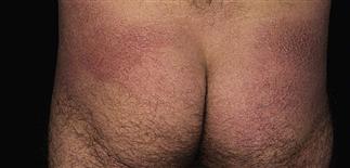
• Parapsoriasis. Controversial term used to describe pre-mycosis fungoides phase. Looks very similar to patch-stage cutaneous T-cell lymphoma (CTCL). May be a precursor stage, but it may not respond to repeated courses of topical steroids, and persist for months or years. • Patch stage. Appears eczematous. Red to pink, scaly, atrophic, mottled, telangiectatic eruption. • Plaque stage. Dusky-red to brown, slightly elevated patches, plaques. Often located on ‘bathing trunk’ area: buttocks, hip, upper thighs. Inner aspects of upper arms, legs (skin folds) involved early. Shape of individual plaques varies: round, oval, arciform, or serpiginous, with central clearing. • Tumor stage. Red-brown expanding nodules, variable in size, may be ulcerated. • Sézary syndrome. Erythroderma and generalized scaling. Palms, soles may be thickened. Alopecia, ectropion common. Infiltration of entire skin produces red, thickened skin with increased scale (exfoliative dermatitis) or without scale (erythroderma). Peripheral node enlargement, generalized pruritus also common.
Frequently misdiagnosed as atopic dermatitis. Other skin diseases resembling CTCL include plaque and pustular psoriasis, drug eruptions, allergic contact dermatitis.
TREATMENT
Stage-related. Referral to dermatologist or oncologist is recommended for staging, treatment. • Patch, plaque stages. Topical chemotherapy (nitrogen mustard, carmustine), psoralen plus ultraviolet A, ultraviolet B, total-body electron beam therapy, interferon, combination of these therapies used. • Tumor stage. Spot radiation, interferon can be effective. • Erythroderma or Sézary syndrome. Extracorporeal photopheresis, interferon, methotrexate, prednisone, cyclophosphamide (Cytoxan), combinations of these therapies used, along with supportive care if needed. • Generally, course and prognosis relate to disease stage and are extremely variable. Early CTCL and patch stages can last many years without progression to tumor development, adenopathy, internal organ involvement. Some patients simply have smoldering inflammatory changes resembling eczema that are kept under control with topical steroids. At the other end of the spectrum is progression to plaque and tumor stage, visceral organ infiltration. Necrosis, ulceration of plaques, tumors common in progressive cases. May eventually involve lymph nodes and viscera; if no response to treatment, CTCL can be fatal.


