Cutaneous manifestations of renal disease

Published on 08/03/2015 by admin

Filed under Dermatology

Last modified 22/04/2025

Print this page

rate 1 star rate 2 star rate 3 star rate 4 star rate 5 star
Your rating: none, Average: 0 (0 votes)

This article have been viewed 2081 times

Chapter 38 Cutaneous manifestations of renal disease

12. What is transepidermal elimination? What is its relationship to kidney disease?

These are a group of diseases in which altered components of skin are eliminated via the epidermis, a process termed transepidermal elimination. Several different perforating diseases have been associated with chronic renal failure, including Kyrle’s disease, reactive perforating collagenosis, and perforating folliculitis. Because features of more than one type of perforating disorder have been noted in skin biopsies from patients with chronic renal failure, it has been suggested that this condition be referred to as the acquired perforating dermatosis of chronic renal failure. This eruption occurs in up to 10% of patients on dialysis but has also developed in patients with renal failure even without dialysis treatment. The lesions consist of keratotic papules and nodules on the trunk and extremities (Fig. 38-2). They occur more commonly in black patients. Skin biopsy confirms the diagnosis. Recently, treatment with narrow band ultraviolet B radiation has been reported to be effective in the treatment of this condition. This eruption may resolve spontaneously over a period of months.

Ohe S, Danno K, Sasaki H, et al: Treatment of acquired perforating dermatosis with narrowband UVB, J Am Acad Dermatol 50:892–894, 2004.

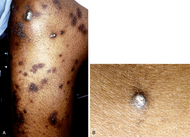
Patterson JW: The perforating disorders, J Am Acad Dermatol 10:561–581, 1984.

14. Describe the porphyria-like eruption of dialysis.

18. How should skin biopsy be used for the diagnosis of systemic amyloidosis?

In primary systemic amyloidosis, immunoglobulin light-chain proteins and serum amyloid P (SAP) are deposited in skin, tongue, heart, spleen, joints, peripheral nerves, and carpal ligaments due to an underlying plasma cell dyscrasia or multiple myeloma. Cutaneous changes may be present, including purpura of the upper trunk, face, and neck and eyelid purpura, which is very characteristic of primary systemic amyloidosis (Fig. 38-3). Waxy papules, particularly on the palms and fingertips, have also been reported. Secondary systemic amyloidosis is due to chronic inflammatory diseases such as tuberculosis and other infections, connective tissue diseases, hidradenitis suppurativa, and familial periodic fever syndromes, and is due to deposition of a distinctive nonimmunoglobulin protein designated AA (amyloid A protein), of which the precursor is an acute-phase reactant produced by the liver. Cutaneous lesions due to amyloid deposits are rarely seen in this type of amyloidosis. Even when there are no cutaneous changes present, skin biopsy may help make a diagnosis of primary or secondary systemic amyloidosis. Clinically, normal skin, abdominal fat, tongue, rectal, and minor salivary gland biopsies have been used to confirm the diagnosis, thus avoiding the need for more invasive biopsies of internal organs.

Wong CK, Wang WJ: Systemic amyloidosis. A report of 19 cases, Dermatology 189:47–51, 1994.