Cutaneous manifestations of AIDS

Published on 05/03/2015 by admin

Filed under Dermatology

Last modified 22/04/2025

Print this page

rate 1 star rate 2 star rate 3 star rate 4 star rate 5 star
Your rating: none, Average: 0 (0 votes)

This article have been viewed 1992 times

Chapter 39 Cutaneous manifestations of AIDS*

3. What are the most common dermatoses associated with HIV infection?

Papulosquamous dermatoses are among the most commonly seen cutaneous manifestations of HIV infection, and these include seborrheic dermatitis (Fig. 39-1) and xerosis. Other common dermatologic conditions include bacterial infections, such as Staphylococcus aureus skin infections. Fungal infections, such as mucocutaneous candidiasis (oropharyngeal and vulvovaginal) and dermatophytosis (tinea pedis, tinea cruris, tinea manuum, and onychomycosis), are also commonly encountered. Frequently seen viral infections include human papilloma virus infections (condyloma acuminata, common and plantar warts), as well as infections with herpes simplex virus, varicella-zoster virus, molluscum contagiosum, and Epstein-Barr virus (oral hairy leukoplakia).

5. What is the most common bacterial pathogen in HIV disease? How does it manifest itself?

Staphylococcus aureus is the most common cutaneous bacterial pathogen in HIV disease. Cutaneous infections due to S. aureus most commonly present as a superficial folliculitis. Less common manifestations include impetigo, ecthyma, furunculosis, cellulitis, abscesses, and botryomycosis. In addition, S. aureus can secondarily infect underlying primary dermatoses such as eczema, scabies, herpetic ulcers, and Kaposi’s sarcoma, or can colonize intravenous catheter sites. Staphylococcal colonization (carriage) of the nose and flexures (perineal, toe webspaces) is known to increase in HIV disease and may account for the increased incidence of cutaneous infections. As in the general population, infections with community-acquired methicillin-resistant S. aureus (MRSA) are becoming increasingly common.

Table 39-1. Mucocutaneous Diseases Seen in HIV Infection*

Neoplastic Diseases Infectious Diseases
Kaposi’s sarcoma
Lymphoma
Squamous cell carcinoma
Basal cell carcinoma
Bacterial
Staphylococcus aureus infections
Syphilis
Bacillary angiomatosis
Fungal
Candida, Penicillium marneffei
Dermatophytosis
Cryptococcosis
Histoplasmosis
Viral
Human papillomavirus (HPV)
Molluscum contagiosum
Herpes simplex virus (HSV)
Varicella-zoster virus (VZV)
Cytomegalovirus (CMV)
Epstein-Barr virus
Arthropods
Scabies
PAPULOSQUAMOUS DISEASES
Seborrheic dermatitis
Xerosis/acquired ichthyosis
Psoriasis
Reiter’s syndrome
MISCELLANEOUS DISEASES
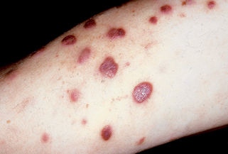
Eosinophilic folliculitis
Drug eruptions
Hyperpigmentations
Photoeruptions
Pruritus
Lipodystrophy
Granuloma annulare
Aphthosis

Dover JS, Johnson RA: Cutaneous manifestations of human immunodeficiency virus infection: parts 1 and 2, Arch Dermatol 127:1383–1391, 1549–1558, 1991.

James W, editor: AIDS: a ten-year perspective, Dermatol Clin 9:391–615, 1991.

* Costner M, Cockerell CJ: The changing spectrum of the cutaneous manifestations of HIV disease, Arch Dermatol 134:1290–1292, 1998.

Ahuja D, Albrecht H: HIV and community-acquired MRSA, AIDS Clin Care 21:21–23, 2009.
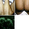
11. What is oral hairy leukoplakia?

Oral hairy leukoplakia, which is predictive for development of AIDS, is primarily seen in HIV-infected patients but also has been described rarely in HIV-negative immunosuppressed organ transplant recipients. It is due to Epstein-Barr virus replication within clinical lesions. Oral hairy leukoplakia occurs primarily on the lateral edges of the tongue as parallel, vertically oriented, white plaques, producing a corrugated appearance (Fig. 39-3A). It can infrequently also involve the dorsal and ventral aspects of the tongue, the buccal or labial mucosa, and the soft palate. The plaque in this condition does not rub off with scraping (unlike candidal thrush) and is usually asymptomatic. Histologically, parakeratosis, acanthosis, and ballooning cells (koilocytes) are seen. In situ Epstein-Barr virus DNA hybridization of lesional scrapings or tissue sections shows positive nuclear staining within epithelial cells. Lesions may respond to acyclovir, zidovudine, podophyllin, tretinoin, or excision but do not respond to anticandidal treatment.

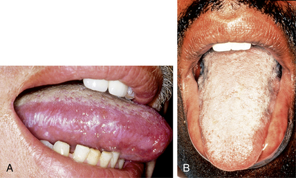
Resnick L, Herbst JS, Raab-Traub N: Oral hairy leukoplakia, J Am Acad Dermatol 22:1278–1282, 1990.

13. What is HIV-associated eosinophilic folliculitis?

HIV-associated eosinophilic folliculitis is a chronic, pruritic dermatosis of unknown etiology characterized by discrete, erythematous, follicular, urticarial papules on the head and neck, trunk, and proximal extremities (Fig. 39-4). Most cases occur in males, but the disease has been reported in females. Bacterial cultures are negative, and the eruption does not resolve with antistaphylococcal treatment. It is associated with peripheral eosinophilia, an elevated serum IgE level, and advanced HIV infection (CD4 counts lower than 250 cells/mm3). Eosinophilic folliculitis is not specific for HIV infection, as it has rarely been described in association with hematologic malignancies.

Transverse histologic sections are superior to vertical sections in the diagnosis of this disease. Histopathologic findings include a perivascular and perifollicular mixed infiltrate with variable numbers of eosinophils and spongiosis of the follicular infundibulum or sebaceous gland with a mixed infiltrate. Treatment options include potent topical corticosteroids, antihistamines, ultraviolet B phototherapy, itraconazole, oral metronidazole, permethrin cream, and isotretinoin.

Piantanida EW, Turiansky GW, Kenner JR, et al: HIV-associated eosinophilic folliculitis: diagnosis by transverse histologic sections, J Am Acad Dermatol 38:124–126, 1998.

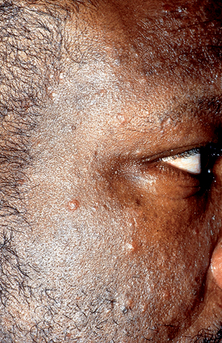
Simpson-Dent SL, Fearfield LA, Staughton RCD: HIV-associated eosinophilic folliculitis: differential diagnosis and management, Sex Transm Infect 75:291–293, 1999.

19. How does varicella-zoster virus infection present in the HIV-positive patient?

20. Do any photosensitive dermatoses occur in HIV disease?

Various photosensitive dermatoses have been described in HIV disease, and these include porphyria cutanea tarda (PCT), lichenoid photoeruptions, and chronic actinic dermatitis. Photosensitivity may, in fact, be the presenting sign of HIV infection.

Most cases of PCT in HIV infection are acquired and many are associated with historical or serologic evidence of hepatitis B or C infection, as well as with elevated transaminase levels and history of alcohol abuse. Patients present with blisters, erosions, crusting, scarring, and increased skin fragility on the face and dorsal hands. In one study, urinary and stool porphyrin excretion patterns classic for PCT occurred in hepatitis C–positive AIDS patients without any clinical evidence of porphyria.

Lichenoid photoeruptions in HIV infection occur most often in black individuals with advanced HIV disease and may be associated with photosensitizing drug use. Patients present with pruritic, violaceous plaques that begin on the face, neck, dorsal hands, and arms. The plaques may become hyperpigmented, hypopigmented, or depigmented and may extend to non–sun-exposed sites. Histopathologic features are primarily those of lichenoid drug eruption or hypertrophic lichen planus, but some patients have findings of lichen nitidus. Patients may improve or clear with discontinuation of a photosensitizing drug, sun avoidance, and sunscreen use.

Chronic actinic dermatitis has been described in markedly immunosuppressed patients and presents as a chronic pruritic and idiopathic eczematous dermatitis in a photodistribution. Phototesting shows increased sensitivity to ultraviolet B. Histologic findings demonstrate eczematous, lymphoma-like, and psoriasiform changes.

O’Connor WJ, Murphy GM, Darby C, et al: Porphyrin abnormalities in acquired immunodeficiency syndrome, Arch Dermatol 132:1443–1447, 1996.

22. Describe some of the potential cutaneous side effects of antiretroviral therapy.

A syndrome of lipodystrophic changes is temporally associated mainly with use of protease inhibitors, and possibly with nucleoside reverse transcriptase inhibitors. It is characterized by enlargement of the dorsocervical fat pad (“buffalo hump”), breast hypertrophy, visceral abdominal fat accumulation (“crix belly,” “protease paunch”) (Fig. 39-5), peripheral fat wasting with prominence of the superficial veins, and loss of fat in the buccal, temporal, and buttocks areas. Lipodystrophic changes have been associated with hypertriglyceridemia, hypercholesterolemia, hyperglycemia, insulin resistance, and hyperinsulinemia. Evidence of associated Cushing’s syndrome or disease is lacking. Lipodystrophic changes have occasionally been reported in patients not taking protease inhibitors. Histologic findings in patients with lipoatrophy include atrophy of the subcutaneous fat, fat lobules with variably sized and often large adipocytes, prominent capillary vascular proliferation, and focal lymphocytic infiltrate and lipogranuloma formation. The exact mechanism involved in these changes is not clear. In addition, antiretroviral therapy has recently been temporally associated with symptomatic angiolipomatosis. Also, painful periungual inflammation (paronychia) of the fingernails and toenails has been reported with use of indinavir and lamivudine. Cutaneous side effects have been described with enfuvirtide (Fuzeon, T-20) use. This drug is a member of a new class of HAART known as fusion inhibitors and is administered subcutaneously. Reported skin side effects are very common and include erythema, induration, nodules, and cysts at the injection sites (Fig. 39-6A,B).

Dank JP, Colven R: Protease inhibitor-associated angiolipomatosis, J Am Acad Dermatol 42:129–131, 2000.

James J, Carruthers A, Carruthers J: HIV-associated facial lipoatrophy, Dermatol Surg 28:979–986, 2002.

Lalezari JP, Henry K, O’Hearn M, et al: Enfuvirtide, an HIV-1 fusion inhibitor, for drug-resistant HIV infection in North and South America, N Engl J Med 348:2175–2185, 2003.

Ward HA, Russo GG, Shrum J: Cutaneous manifestations of antiretroviral therapy, J Am Acad Dermatol 46:284–293, 2002.