74
Cutaneous fungal
infections (tinea)
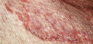
A very characteristic pattern of inflammation is the active border of infection.
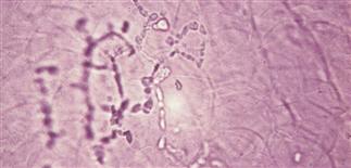
Dermatophytes appear as translucent, branching, rod-shaped filaments (hyphae) of uniform width, with lines of separation (septa) spanning the width and appearing at irregular intervals.
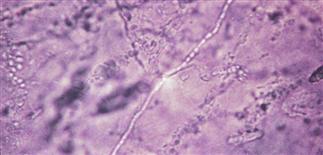
Hyphae may be difficult to find in a potassium hydroxide wet mount. Parker’s blue ink and other stains easily stain the hyphae, rendering them visible under low power.
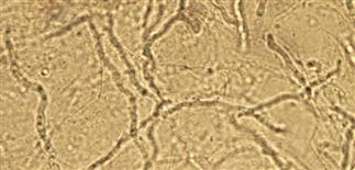
Mosaic artifact. Lipid droplets between epithelial cells simulate fungal hyphae in potassium hydroxide wet mounts. Heat will make this disappear.
DESCRIPTION
Tinea is the clinical term for dermatophyte infection. Dermatophytes can infect and survive only on dead keratin; that is, the top layer of the skin (stratum corneum), hair, nails.
HISTORY
Some species are able to infect the hair shaft in addition to the skin.
• Endothrix pattern: fungal hyphae inside the hair shaft. • Ectothrix pattern: fungal hyphae inside and on the surface of the hair shaft.
Fungal spores are either large or small. The type of hair invasion is classified as large-spored or small-spored ectothrix, and large-spored endothrix.
PHYSICAL FINDINGS
• Dermatophytes produce various disease patterns that vary with location: foot (tinea pedis), groin (tinea cruris), body (tinea corporis), face (tinea faciei), hand (tinea manuum), scalp (tinea capitis), beard (tinea barbae), nails (onychomycosis). • Greatest numbers of hyphae located in active border, which is best area to get a sample for potassium hydroxide examination. Active border is scaly, red, slightly elevated. Hold a #15 surgical blade perpendicular to skin surface; smoothly draw the blade against the scale. Place scale on microscope slide and apply coverslip. Potassium hydroxide (10% solution) is applied to edge of coverslip and allowed to run under.
• With the microscope condenser moved down away from the slide, scan entire area under coverslip at × 10 power. Dermatophytes appear as translucent, branching, rod-shaped filaments (hyphae) of uniform width, with lines of separation (septa) spanning width and appearing at irregular intervals. Uniform width and branching distinguish hyphae from hair and other debris. • Fungal culture necessary for hair and nail fungal infections. Cultures usually become positive in 1–2 weeks. Three types of culture media used for tinea. Sabouraud’s agar: allows growth of most fungi, including non-dermatophytes; useful for nail infections, because detection of non-dermatophytes is desirable. Mycosel agar: contains antibiotics to prevent growth of bacteria and saprophytic fungi; best for evaluation of hair tinea. Dermatophyte test medium: supplied in vial kits, produces fast but slightly less accurate results; yellow medium turns pink in presence of dermatophytes in 6–7 days.
TREATMENT
Topical or systemic depending on site, extent of involvement. Generally, hair and nail infections require longer treatment. Specific sites are covered in the chapters that follow.


