103 Consolidation
Questions
How would you investigate suspected bacterial pneumonia?
• FBC, serum urea, electrolytes and liver function tests
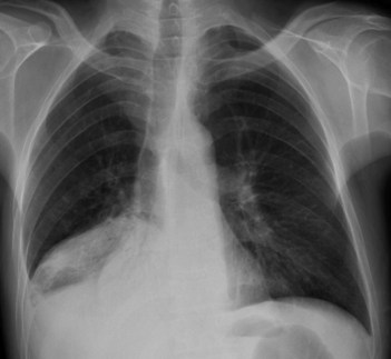
• Chest radiography (Fig. 103.1)
• Test for Legionella (culture, direct fluorescent-antibody test or urinary antigen assay), mycoplasma immunoglobulin M
• Consider serological testing for HIV (for patients 15–54 years, particularly when there is lymphopenia or a low CD4 cell count).
Advanced-level questions
How do you determine the severity?
Using the CURB65 severity score, 1 point for each feature present:
The prognosis based on CURB65 score:
4 or 5: assess with specific consideration to the need for transfer to a critical care unit
2: moderate risk of death; considered for short-stay inpatient treatment or hospital-supervised outpatient treatment
0 or 1: low risk of death; may be suitable for therapy at home.
The CURB65 severity score should be interpreted in context of the overall clinical picture.
What are the extrapulmonary manifestations of mycoplasma pneumonia?
Which antibiotics would you use in a patient with community-acquired pneumonia where the pathogen is not known?
What are the poor prognostic factors in patients with community-acquired pneumonia?
• Coexisting conditions such as cardiac failure, renal failure, COPD, malignancy
• Clinical features: respiratory rate >30 breaths/min, hypotension (systolic BP <90 mmHg or diastolic BP <60 mmHg), temperature >38.3°C, impaired mental status (stupor, lethargy, disorientation or coma), extrapulmonary infection (e.g. septic arthritis, meningitis)
• Investigations: haematocrit <30%, white cell count <4 or >30 × 106 cells/l, azotaemia, arterial blood gas <60 mmHg while breathing room air, chest radiograph showing multiple lobe involvement, or rapid spread or pleural effusion
What do you know about pulmonary eosinophilic disorders?
Löffler syndrome. This is characterized by transient pulmonary infiltrates and peripheral eosinophilia. It is associated with parasitic infections, drug allergies and exposure to inorganic chemicals such as nickel carbonyl. The course is benign and respiratory failure almost unknown.
Eosinophilia in asthmatics. The most common cause is allergic bronchopulmonary aspergillosis. This condition is benign but chronic.
Tropical eosinophilia. This is secondary to filarial infection (Wuchereria bancrofti or W. malayi brugi).
Churg–Strauss syndrome. Diagnosis requires four of the following features: asthma; eosinophilia >10%, mononeuropathy or polyneuropathy; paranasal sinus abnormality; non-fixed pulmonary infiltrates visible on chest radiographs; and blood vessels with extravascular eosinophils found on biopsy.
Chronic eosinophilic pneumonia. This chronic debilitating illness is characterized by malaise, fever, weight loss and dyspnoea. The chest radiograph shows a peripheral alveolar filling infiltrate predominantly in the upper lobes (the ‘photographic negative’ of pulmonary oedema).
What is the pathogenesis of ventilator-acquired pneumonia?
Hospital-acquired pneumonia. Micro-aspiration is the primary route of bacterial entry into the lower respiratory tract. Risk factors include sedation, intubation for operative procedures, vomiting and impaired swallowing.
Ventilator-associated pneumonia. Leakage of bacteria and oral secretions around the endotracheal cuff, inhalation of contaminated aerosols, or reflux of contaminated ventilator tubing condensate are the primary routes of bacterial entry into the lower respiratory tract. Promising biomarkers include procalcitonin, C-reactive protein and soluble triggering receptor expressed on myeloid cells (sTREM-1).
Outcomes. The ‘battlefield’ between the pathogens entering the lower respiratory tract and the host defences determines possible outcomes: colonization, tracheobronchitis or hospital/ventilation-acquired pneumonia.


