Connective tissue diseases
The inflammatory disorders of connective tissue often affect several organs, as in systemic lupus erythematosus (LE), but they may also involve the skin alone (e.g. discoid LE). Autoantibodies are a feature of these diseases, which can thus be regarded as ‘autoimmune’.
Lupus erythematosus
Clinical presentation
Systemic lupus erythematosus (99% ANA, 50% Ro, 60% dsDNA positive)
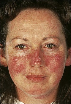
Skin signs are found in 80%. The facial butterfly eruption (Fig. 1) is characteristic, but photosensitivity, discoid lesions, diffuse alopecia, mouth lesions and vasculitis also occur. Multisystem involvement with serological or haematological abnormalities must be demonstrated to diagnose systemic LE (Table 1). The female : male ratio is 8 : 1.
| Organ | Involvement |
|---|---|
| Skin | Photosensitivity, facial rash, vasculitis, hair loss, Raynaud’s phenomenon |
| Blood | Anaemia, thrombocytopenia |
| Joints | Arthritis, tenosynovitis, calcification |
| Kidney | Glomerulonephritis, nephrotic syndrome |
| Heart | Pericarditis, endocarditis, hypertension |
| Central nervous system | Psychosis, infarction, neuropathy |
| Lungs | Pneumonitis, effusion |
Discoid lupus erythematosus (35% ANA, 2% Ro, <5% dsDNA positive)
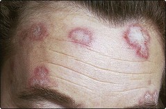
One or more round or oval plaques appear on the face, scalp or hands (Fig. 2). The lesions are well demarcated, red, atrophic, scaly and show keratin plugs in dilated follicles. Scarring leaves alopecia on the scalp and hypopigmentation in those with a pigmented skin. Remission occurs in over 50%. Internal involvement is not a feature, and only 6% develop systemic LE. Women outnumber men by 2 : 1.
Systemic sclerosis
Clinical presentation
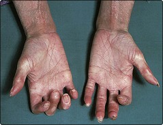
Raynaud’s phenomenon (p. 70) is frequently the presenting sign. The skin of the fingers, forearms and lower legs becomes tight, waxy and stiff, and the finger pulps are resorbed (Fig. 3). Facial signs include perioral furrowing, telangiectasia (p. 115) and restricted mouth opening. Internal organ involvement, e.g. renal failure, may prove fatal (Table 2). Women are affected more than men (F : M ratio 4 : 1). Some 90–95% of cases are ANA positive. The diagnosis is rarely in doubt in diffuse disease, although chronic graft-versus-host disease shows similar changes. Limited cutaneous systemic sclerosis has a better prognosis and usually affects only the neck, forearms and lower legs. A subset described as CREST syndrome, is confined to Calcinosis, Raynaud’s phenomenon, oEsophageal dysmotility, Sclerodactyly and Telangiectasia.
| Organ | Involvement |
|---|---|
| Skin | Raynaud’s phenomenon, calcinosis, sclerodactyly, telangiectasia |
| Gut | Oesophageal dysmotility, malabsorption, dilated bowel |
| Lung | Fibrosis, pulmonary hypertension |
| Heart | Pericarditis, myocardial fibrosis |
| Kidney | Renal failure, hypertension |
| Muscle | Myositis, tendon involvement |
Management
Treatment is mainly supportive. Nifedipine can help Raynaud’s phenomenon. Hypertension is controlled. Systemic steroids, penicillamine and immune-suppressives have been used with little benefit. Photophoresis may be tried (p. 115). Renal crises (associated with antiRNA polymerase I and III antibodies) should be managed aggressively with angiotensin converting enzyme (ACE) inhibitors.
Morphoea (localized scleroderma)
Morphoea presents with round or oval plaques of induration and erythema, often with a purplish edge (Fig. 4). These become shiny and white, eventually leaving atrophic hairless pigmented patches. The trunk or proximal limbs are affected. Morphoea is more common in women (F : M ratio 3 : 1). Linear morphoea may involve the face or a limb and, when seen in a child, can retard growth of the underlying tissues, including bone.
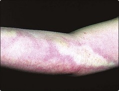
Fig. 4 Morphoea.
Seen here on the arm of a child. The white indurated plaque has an erythematous edge.
There is no well established treatment (p. 115), although topical steroids are often given. The disease usually resolves spontaneously within months to years.
Dermatomyositis
Clinical presentation
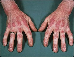
In dermatomyositis/polymyositis, skin changes or muscle weakness may predominate. The typical eruption is a lilac–blue discoloration around the eyelids, cheeks and forehead, often with oedema. Bluish–red papules or streaks on the dorsal aspects of the hands (Fig. 5), elbows and knees are seen, sometimes with pigmentation and nail fold telangiectasia. Photosensitivity is common. There is no strong association with autoantibodies, although anti-Jo-1 antibodies predict lung involvement. An association with malignancy exists in patients over 40 years, 40% of whom have an underlying tumour, usually of the lung, breast or stomach. A childhood variant mainly affects the muscles and causes calcinosis and contractures.
Management
Investigations must define the degree of myositis and, in adults, exclude the possibility of underlying neoplasia. Treatment is with systemic steroids in moderate to high dosage, often with an immunosuppressive such as azathioprine or methotrexate. Immunoglobulin infusion and possibly photophoresis (p. 115) may help.
Connective tissue diseases
 Systemic LE is an autoimmune, multisystem disease in which a butterfly rash, photosensitivity, vasculitis and alopecia may be seen. Treatment depends on the type and degree of involvement and often includes systemic steroids and immunosuppressive agents.
 Subacute LE is a less aggressive form of LE with predominantly cutaneous features.


