139
Congenital melanocytic nevi
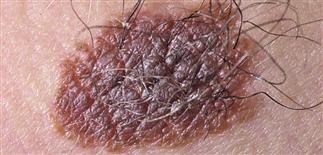
Congenital nevi are benign skin tumors composed of melanocyte-derived nevus cells. Many lesions have a pebbled surface and course terminal hair.
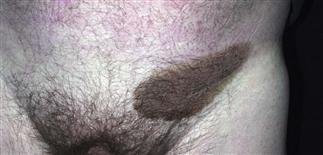
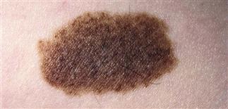
Congenital nevi with uniform surface characteristics and light pigmentation have little chance of degenerating into melanoma.
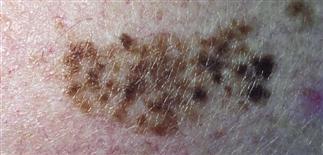
Congenital nevus with speckled surface showing uniform, darkly pigmented papules on a lightly pigmented background.
Congenital nevi may have a papular surface that mimics cobblestones. Hyperpigmented brown papules are uniformly distributed over the surface.
DESCRIPTION
Benign growths composed of melanocyte-derived nevus cells, present at birth or appearing by age 2 years. Considered a type of birthmark. Many variants.
HISTORY
• Any melanocytic nevus present at birth or appearing during infancy is considered a congenital melanocytic nevus. Roughly 1% of newborns have at least one. • Nevi may increase in size, become more heavily pigmented during puberty. • Usually asymptomatic but may be irritated by clothing, external trauma.
PHYSICAL FINDINGS
• Usually brown, raised, with an irregular verrucous surface. Most have increased terminal hairs. Size varies greatly. Depending on their location, large lesions may be disfiguring. • Mongolian spots are poorly defined patches colored blue-black to gray, more commonly seen in the sacral region of newborns of darker skin. • Congenital nevi are usually compound nevi with nevus cells at the junction and in the dermis. Nevus cells may extend into fat, investing adnexal structures and blood vessels. • Mongolian spots are equivalent to blue nevi histologically, with pigmented spindle-shaped nevus cells deep in the dermis. • Most congenital melanocytic nevi are benign and follow the usual course of maturation. Nevi that deviate from this pattern are suspicious, and biopsy is warranted. Mongolian spots often fade in early childhood. • The risk of malignant degeneration occurring in congenital melanocytic nevi is controversial. There is general agreement that risk of malignant change is increased in congenital nevi with diameters greater than 20 cm. Lifetime risk is estimated at 5–8%. The risk of melanoma developing in smaller congenital nevi is uncertain but is likely increased in lesions larger than 2 cm. • Many dermatologists favor elective excision of congenital nevi when feasible, usually around the time of puberty. Careful histologic review is needed by a qualified dermatopathologist.
TREATMENT
• Assess all the patient’s nevi. • Educate patients and parents on how to perform self-examination of the skin; encourage them to do so on a regular basis. Combine the teaching of self-examination techniques with the screening skin examination. Review changes to watch for, including symptoms of itching and tenderness, with the patient. • Benign nevi are usually symmetric with well-defined borders, uniform in color. Congenital nevi should increase in size in proportion to the patient’s growth through adolescence. Pigmentation should remain uniform. • Photography helpful for following nevi. • Biopsy suspicious nevi. • Large congenital nevi may be disfiguring, depending on location. Plastic surgery referral should be considered for giant congenital nevi. Removal may require serial procedures with tissue expanders.


