Congenital Anomalies and Benign Conditions of the Ovaries and Fallopian Tubes

Published on 10/03/2015 by admin

Filed under Obstetrics & Gynecology

Last modified 22/04/2025

Print this page

rate 1 star rate 2 star rate 3 star rate 4 star rate 5 star
Your rating: none, Average: 0 (0 votes)

This article have been viewed 3886 times

Chapter 20 Congenital Anomalies and Benign Conditions of the Ovaries and Fallopian Tubes

The ovaries and fallopian tubes, along with the blood vessels, ligaments, and connective tissues located along both sides of the uterus, are referred to as the adnexa. In this chapter, the normal development of the ovaries and fallopian tubes is discussed to facilitate a better understanding of the workup and management of benign adnexal masses.

image Congenital Anomalies of the Ovaries and Fallopian Tubes

Abnormal embryologic development of the ovaries is uncommon. Congenital duplication or absence of ovarian tissue may occur, as may ectopic ovarian tissue and supernumerary ovaries. Although rare, the sexual bipotentiality noted in embryologic development can progress without the usual regression of one system, producing an ovotestis and subsequent intersex problems.

Genetic chromosomal disorders, such as Turner syndrome (45 XO), are associated with a lack of normal gonadal development, as evidenced by the rudimentary streaked ovaries that are a hallmark of the disorder. Women with Turner syndrome usually progress through puberty and develop secondary sexual characteristics but enter menopause shortly thereafter. This provides evidence that two X chromosomes are required for normal ovarian development. Testicular predominance occurs with the addition of a single Y chromosome, even in the face of multiple X chromosomes. Such predominance is seen in Klinefelter syndrome (47 XXY), in which testicular development occurs embryologically. In complete androgen insensitivity syndrome (46 XY), which is also known as testicular feminization, the lack of androgen receptors produces a phenotypic female in the face of a Y chromosome. The gonads in these women (functioning testes) should be removed (usually after puberty) because of their significant malignant potential.

Isolated anomalies of the fallopian tubes, the end result of abnormal development of the proximal unfused portions of the paramesonephric ducts, are rare. Aplasia or atresia, usually of the distal ampullary segment of the fallopian tube, is most commonly unilateral in the presence of otherwise normal development. Bilateral aplasia is noted in some cases of uterine and vaginal agenesis. Complete duplication of the fallopian tubes is rarely seen, but distal duplication and accessory ostia are relatively common.

In addition, women exposed in utero to certain drugs, such as diethylstilbestrol (DES), may have abnormalities in the architecture of the fallopian tubes; with DES exposure, the tubes may be shortened, distorted, or clubbed.

image Benign Conditions of the Ovaries

FUNCTIONAL AND BENIGN OVARIAN TUMORS

The human ovary has a striking propensity to develop a wide variety of tumors, most of which are benign. As indicated in Table 20-1, ovarian masses may be functional, inflammatory, metaplastic, or neoplastic. During the childbearing years, 70% of noninflammatory benign ovarian tumors are functional. The remainder are either neoplasms (20%) or endometriomas (10%). The management of ovarian tumors, whether functional, benign, or malignant, involves difficult decisions that may affect a woman’s hormonal status or her future fertility. Although only functional cysts and benign ovarian neoplasms are considered in detail in this chapter, diagnostic methods to differentiate benign masses from malignant ones are discussed.

TABLE 20-1 DIFFERENTIAL DIAGNOSIS OF OVARIAN MASSES

Pathogenesis Specific Type
Functional Follicular cysts
  Lutein cysts
  Polycystic ovaries
Inflammatory Salpingo-oophoritis
  Pyogenic oophoritis—puerperal, abortal, or related to an intrauterine device
  Granulomatous oophoritis
Metaplastic Endometriomas
Neoplastic Premenarchal years—10% are malignant
  Menstruating years—15% are malignant
  Postmenopausal years—50% are malignant

Functional Ovarian Cysts and Tumors

Dozens of ovarian follicles form the “cohort of follicles” of each menstrual cycle. To be classified a functional cyst, the follicle must reach a diameter of at least 3 cm. Functional cysts may cause pelvic pain, a dull sensation, or heaviness in the pelvis.

A follicular cyst, lined by one or more layers of granulosa cells, develops when an ovarian follicle fails to rupture. Similarly, a lutein cyst may develop if the corpus luteum becomes cystic, grows to larger than 3 cm, and fails to regress normally after 14 days. Hemorrhagic cysts, especially hemorrhagic corpus luteum cysts, are more likely to cause symptoms and are more vulnerable to rupture toward the end of the menstrual cycle. The hemorrhage within the cyst results from invasion of the ovarian vessels into the corpus luteum cyst 2 to 3 days after ovulation.

Other specific types of lutein cysts may occur with abnormally high serum levels of human chorionic gonadotropin (hCG) or increased ovarian sensitivity to gonadotropins. Theca-lutein cysts may develop in association with the high levels of hCG present in patients with a hydatidiform mole or choriocarcinoma. Patients undergoing ovulation induction with gonadotropins or clomiphene may also develop theca-lutein cysts. Theca-lutein cysts are usually bilateral, may become quite large (>30 cm), and characteristically regress slowly after the gonadotropin level falls. Rarely, when follicles are stimulated with gonadotropins, theca-lutein cysts can become so extensive as to cause massive ascites and dangerous problems with systemic fluid imbalance.

A luteoma of pregnancy is a related condition in which there is a hyperplasic reaction of ovarian theca cells, presumably from prolonged hCG stimulation during pregnancy. The luteomas characteristically appear as brown to reddish-brown nodules that may be cystic or solid. A luteoma of pregnancy (Figure 20-1) may be associated with multifetal pregnancies or hydramnios. They can cause maternal virilization in 30% of women and, less often, ambiguous genitalia in a female fetus. Although ovarian enlargement may be impressive, surgical resection is not indicated because luteomas regress spontaneously postpartum.

image

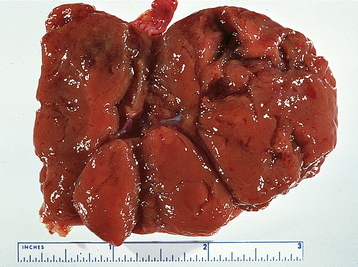
FIGURE 20-1 Gross appearance of a luteoma of pregnancy. Note the multiple brown nodules.

(From Voet RL: Color Atlas of Obstetric and Gynecologic Pathology. St. Louis, Mosby, 1997.)

Polycystic ovary syndrome, a functional disorder generally associated with chronic anovulation and hyperandrogenism, can also produce enlarged ovaries with multiple simple follicles (Figure 20-2). The hormonal aspects of this syndrome are discussed further in Chapter 32.

image

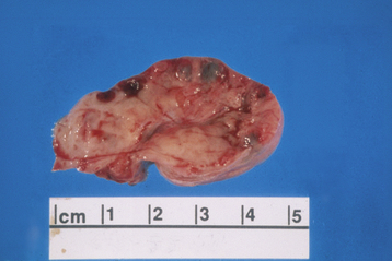
FIGURE 20-2 Ovary with multiple cysts lining the capsule consistent with polycystic ovary syndrome.

(Courtesy of Dr. Sathima Natarajan, Ronald Reagan–UCLA Medical Center.)

C. SERUM CA-125 TITER Absolute value

Risk of Malignancy Index = A × B × C. A cut-off value of 200 discriminates a benign from a malignant mass with a sensitivity of 87% and a specificity of 97%.

Data from Jacobs I, Oram D, Fairbanks J, et al: A risk of malignancy index incorporating CA 125, ultrasound and menopausal status for the accurate preoperative diagnosis of ovarian cancer. Br J Obstet Gynaecol 97:922, 1990.

When a patient has delayed menses, abnormal uterine bleeding, or severe pelvic pain, the differential diagnosis must include ectopic pregnancy, pelvic abscess, or adnexal torsion of a neoplastic cyst. A pregnancy test (hCG), diagnostic laparoscopy, or rarely, laparotomy may be needed. Table 20-3 lists the current modalities that are available to evaluate adnexal masses along with the sensitivity and specificity as calculated by the U.S. Agency for Healthcare Research and Quality. None of these modalities is accurate enough to be used alone for diagnosis.

TABLE 20-3 MODALITIES FOR THE EVALUATION OF ADNEXAL MASSES

Modality Sensitivity (%) Specificity (%)
Gray-scale transvaginal ultrasonography 0.82-0.91 0.68-0.81
Doppler ultrasonography 0.86 0.91
Computed tomography 0.90 0.75
Magnetic resonance imaging 0.91 0.88
Positron emission tomography 0.67 0.79
CA-125 level measurement 0.78 0.78

Data from Agency for Healthcare Research and Quality (AHRQ): Management of adnexal mass. AHRQ Publication No. 06-E004. Rockville, MD, AHRQ, 2006.

Benign Neoplastic Ovarian Tumors

Ovarian neoplasms may be divided generally by cell type of origin into three main groups: epithelial, stromal, and germ cell. Taken as a group, the epithelial tumors are by far the most common, although the single most common benign ovarian neoplasm is the benign cystic teratoma (dermoid cyst), which is a germ cell tumor.

EPITHELIAL OVARIAN NEOPLASMS

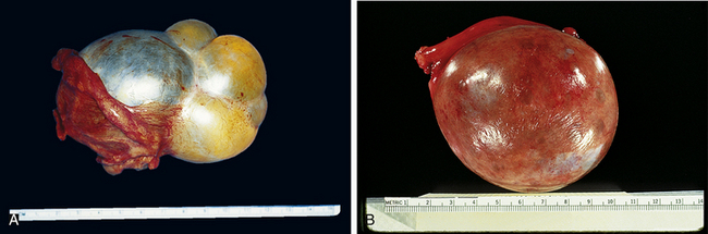
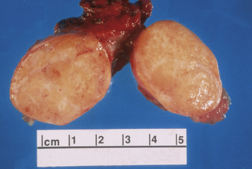
These tumors are believed to be derived from the mesothelial cells lining the peritoneal cavity and also lining the surface of the ovary. The mucinous ovarian neoplasm cytologically resembles the endocervical epithelium, the endometrioid neoplasm resembles the endometrium, and serous tumors resemble the lining of the fallopian tubes. The most common epithelial ovarian tumors are serous cystadenomas. Figure 20-3 shows the gross appearance of a mucinous and serous cystadenoma.

Each of the epithelial ovarian neoplasms has characteristic clinical and histologic features. The serous tumors are bilateral in about 10% of cases. Of all serous tumors, about 70% are benign, 5% to 10% have borderline malignant potential, and 20% to 25% are malignant, depending largely on the patient’s age. Larger serous cystadenomas tend to be multilocular, although small unilocular serous cystomas also occur. Histologically, serous tumors characteristically form psammoma bodies (from the Greek psammos, meaning sand), which are calcific, concentric concretions. Psammoma bodies occur occasionally in benign serous neoplasms and frequently in serous cystadenocarcinomas. Papillary patterns are also common.

The mucinous neoplasms of the ovary can attain a huge size, often filling the entire pelvis and abdomen. They are often multilocular, and benign mucinous tumors are bilateral in less than 10% of cases. About 85% of mucinous tumors are benign. Mucinous tumors are often associated with a mucocele of the appendix. Rarely, a benign mucinous tumor may be complicated by pseudomyxoma peritonei, a condition in which a great many benign implants are seeded onto the surface of the bowel and other peritoneal surfaces and produce large quantities of mucus.

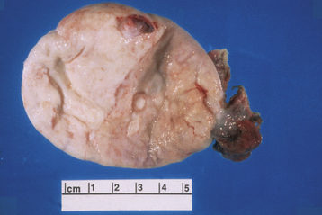
The Brenner tumor is a small, smooth solid ovarian neoplasm, usually benign, with a large fibrotic component that encases epithelioid cells that resemble transitional cells of the bladder (Figure 20-4). In about 33% of cases, Brenner tumors are associated with mucinous epithelial elements.

image

FIGURE 20-4 Gross appearance of a cut-open Brenner tumor.

(Courtesy of Dr. Sathima Natarajan, Ronald Reagan–UCLA Medical Center.)

SEX CORD–STROMAL OVARIAN NEOPLASMS

These tumors include fibromas, granulosa-theca cell tumors, and Sertoli-Leydig cell tumors. Combinations of the latter two types are termed gynandroblastomas.

The tumors in this category derive from the sex cords and specialized stroma of the developing gonad. The embryologic origins of granulosa and theca cells, as well as their counterparts in the testes, the Sertoli and Leydig cells, arise from cells that make up this specialized gonadal stroma. If the ultimate differentiation of cell types occurring in the tumor is feminine, the neoplasm becomes a granulosa cell tumor, a theca cell tumor, or in many instances, a mixed granulosa-theca cell tumor. Neoplasms containing cells that take on a masculine differentiation become Sertoli-Leydig cell tumors. This is far less common. The fibroma represents a stromal cell neoplasm developing from mature fibroblasts in the ovarian stroma.

The granulosa-theca cell neoplasms, as well as their androgenic counterparts, are generally referred to as functioning (not functional) ovarian tumors. They occur in any age group, from birth on, but more commonly in the postmenopausal years. Their functioning characteristics are responsible for a variety of associated presenting signs and symptoms. The granulosa-theca cell tumors promote feminizing signs and symptoms, such as precocious menarche, precocious thelarche, or premenarchal uterine bleeding during infancy and childhood. In the reproductive years, menorrhagia (with alternating amenorrhea), endometrial hyperplasia, and not infrequently, endometrial cancer, breast tenderness, and fluid retention occur. Postmenopausal bleeding may occur in older women with granulosa-theca cell tumors. In contrast, the less frequent Sertoli-Leydig cell tumors are responsible for virilizing effects, such as hirsutism, temporal baldness, deepening of the voice, clitoromegaly, and a defeminizing change in body habitus to a muscular build. Fifteen percent of these tumors produce no obvious endocrinologic effects. Except for the pure thecoma, all these tumors have low malignant potential and are discussed further in Chapter 39.

The ovarian fibroma, another ovarian stromal tumor, forms a solid, encapsulated, smooth-surfaced tumor made up of interlacing bundles of fibrocytes. It is not hormonally active. It is glistening white on its cut surface (Figure 20-5), as opposed to the soft yellow appearance of the granulosa-theca cell or the smaller hilus cell tumor. On occasion, this tumor is associated with ascites caused by the transudation of fluid from the ovarian fibroid. The flow of this ascitic fluid through the transdiaphragmatic lymphatics into the right pleural cavity may result in Meigs’ syndrome (ascites and hydrothorax in association with an ovarian fibroma). The ovarian fibroma may be associated with theca cell elements called a fibrothecoma.

image

FIGURE 20-5 Gross appearance of an ovarian fibroma.

(Courtesy of Dr. Sathima Natarajan, Ronald Reagan–UCLA Medical Center.)

GERM CELL TUMORS

Germ cell neoplasms can occur at any age. They make up about 60% of ovarian neoplasms occurring in infants and children.

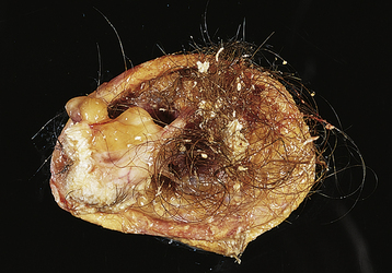
The most common ovarian neoplasm is the benign cystic teratoma, a germ cell tumor that can take on a great variety of forms, with virtually all adult tissues being represented within the mass. Ten percent to 15% of teratomas are bilateral. The benign cystic teratoma, commonly referred to as a dermoid cyst, is composed primarily of ectodermal tissue (such as sweat and sebaceous glands, hair follicles, and teeth), with some mesodermal and rarely endodermal elements. These are slow-growing tumors. Half are diagnosed in women between 25 and 50 years of age. Most are less than 10 cm in diameter. Because of the oily secretion of the sebaceous glands, the desquamated squamous cells, the presence of hair, and the presence of a dermoid tubercle (of Rokitansky), which often contains a hard, well-formed tooth, the dermoid cyst has a characteristic gross and histologic appearance (Figure 20-6). Other tissue components commonly found in benign cystic teratomas include mature brain, bronchus, thyroid, cartilage, intestine, bone, and carcinoid cells. As opposed to similar tissues found in a malignant immature teratoma, the tissues making up the benign (mature) teratoma are all of an adult, well-differentiated form.

image

FIGURE 20-6 Gross appearance of a cut-open dermoid cyst. Note the presence of hair-bearing skin.

(From Voet RL: Color Atlas of Obstetric and Gynecologic Pathology. St. Louis, Mosby, 1997.)

DIAGNOSIS OF BENIGN OVARIAN TUMORS

The clinical features of benign ovarian tumors are often nonspecific. Except for the functioning ovarian neoplasms, most benign ovarian tumors are asymptomatic unless they undergo torsion or rupture. They usually enlarge very slowly, so that an increase in abdominal girth or pressure on surrounding organs is not perceived until the later stages of growth. Any pelvic pain is generally mild and intermittent, unless the tumor twists on its pedicle (torsion), when infarction may induce severe pain and tenderness. On rare occasions, an ovarian cyst may rupture spontaneously from internal hemorrhage or intracystic pressure, resulting in pain and peritoneal irritation. A cyst may also rupture occasionally during or after a bimanual pelvic examination or with intercourse. Depending on the cystic contents, pain of varying degrees of severity can result. The escape of thin serous fluid without hemorrhage may evoke little pain or tenderness, but the oily contents of a dermoid cyst or the thick mucinous fluid of a mucinous cystadenoma may be irritating to both the parietal and the visceral peritoneum, with the development of chemical peritonitis and possibly the subsequent formation of troublesome intraabdominal adhesions.

Bimanual pelvic examination generally indicates the presence of the tumor in the pelvis, but the tumor may be too small to be palpated. On the other hand, if the tumor is large enough, it may be detected by abdominal palpation. Examination may suggest a cystic mass or a solid tumor. Movement of the mass separate from the uterus supports the suspicion of an adnexal mass instead of a uterine leiomyoma. Percussion of the abdomen in a patient with a large ovarian cyst reveals dullness anteriorly with tympany in the flanks as the bowel is displaced laterally by the tumor.

If the tumor undergoes torsion and infarction or rupture, signs of peritoneal irritation may be present. If complete infarction has occurred, there may be abdominal rigidity. Paralytic ileus may also be present.

Pelvic ultrasonography, particularly transvaginal ultrasonography, with or without color Doppler, may be helpful. A pelvic ultrasound will be highly suggestive of a dermoid cyst, especially if it is found to include a tooth-like calcification.

Tumor markers, such as serum CA 125, as part of the RMI (see Table 20-2) may help to distinguish between benign and malignant masses, particularly in a postmenopausal patient. When clinical evaluation, pelvic ultrasonography, and tumor markers all indicate malignancy, the positive predictive value of the combination is high in postmenopausal women. Such patients should be referred to a gynecologic oncologist for surgical evaluation.

Laparoscopy is helpful in distinguishing between a uterine myoma, a quiescent hydrosalpinx, and an ovarian tumor, but it will not distinguish between a functional cyst, a benign neoplasm, and an encapsulated malignant ovarian neoplasm. On occasion, laparoscopy may identify endometriosis on the surface of the ovary. An ovarian endometrioma cannot be distinguished unequivocally from an ovarian neoplasm by visualization alone. In general, laparotomy is preferable to laparoscopy in the ultimate evaluation of a suspicious adnexal mass unless the entire mass can be removed laparoscopically without rupture for histologic examination.

Management of Ovarian Neoplasms

No persistent ovarian neoplasm should be assumed to be benign until proved so by surgical exploration and pathologic examination. The indications for exploratory laparotomy in a patient with a pelvic mass have been discussed under functional tumors. If laparotomy is indicated, any ascitic fluid should be collected on opening the peritoneal cavity and sent for cytologic examination. A frozen-section histologic diagnosis should be obtained intraoperatively to exclude malignancy. The definitive treatment will depend on the type of neoplasm, the patient’s age, and her desire for future childbearing.

Benign epithelial ovarian neoplasms are generally treated by unilateral salpingo-oophorectomy. The contralateral ovary must be carefully inspected to exclude a bilateral lesion. Because of the possible coexistence of an appendiceal mucocele with a mucinous cystadenoma, appendectomy should also be performed in such patients. If the patient is young and nulliparous, the ovarian neoplasm is unilocular, and there are no excrescences within the cyst, an ovarian cystectomy with preservation of the ovary may be performed. In an older woman, a total abdominal hysterectomy and bilateral salpingo-oophorectomy may be appropriate, particularly if there is any suspicion of malignancy.

Stromal cell neoplasms of the ovary are generally treated by unilateral salpingo-oophorectomy when future pregnancies are a consideration. Ovarian fibromas, even when associated with ascites and a right hydrothorax (Meigs’ syndrome), are almost always benign and might even be treated by resection from the ovary in a young woman.

Cystic teratomas (“dermoids”) can be treated by ovarian cystectomy. Because 15% to 20% are bilateral, the contralateral ovary should be carefully evaluated and any cysts resected.

In a patient with a gonadoblastoma, dysgenetic ovaries are usually present, necessitating bilateral salpingo-oophorectomy, particularly in the presence of a Y chromosome. With embryo transfer now available to these patients, the uterus should be left in situ if future fertility is desired.

image Benign Conditions of the Fallopian Tubes

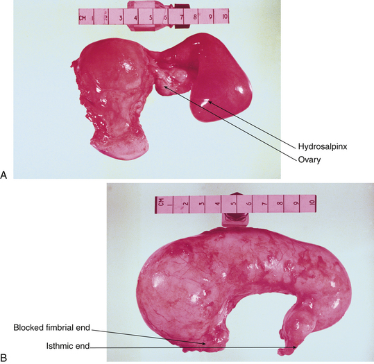
Most benign “tumors” of the fallopian tubes are infectious or inflammatory (e.g., hydrosalpinx and pyosalpinx [Figure 20-7]). Benign neoplasms of the oviducts are rare. Although the tubes, uterine corpus, and uterine cervix are from the same müllerian anlage (primordial tissue), the tubes have less of a tendency toward neoplastic transformation.

Tubal neoplasms that do occur are epithelial adenomas and polyps, myomas from the tubal musculature, inclusion cysts from the mesothelium, or angiomas from the tubal vasculature.

It is quite difficult to differentiate a tubal neoplasm from other adnexal masses on examination, and operative exploration is generally necessary to confirm the diagnosis. Salpingectomy represents the definitive treatment, although if pathologic evaluation confirms the benign nature of the neoplasm, normal portions of the tube may be preserved for fertility reasons in selected cases.

As the name parovarian (beside the ovary) implies, parovarian neoplasms are generally located within the broad ligament between the tube and the ovary. These tumors are generally small compared with ovarian cysts, measuring less than 8 cm in diameter. Histologically, most appear to be derived from paramesonephric (müllerian) structures or occasionally from mesonephric (wolffian) remnants. Although the malignancy rate is less than 10%, it is necessary to resect the cystic mass to obtain a pathologic assessment.

Torsion either of the ovary alone or of both the ovary and fallopian tube (adnexal torsion) represents an acute surgical emergency. Torsion is a complication of benign ovarian tumors, parovarian cysts, and tubal ligation remnants. Adnexal torsion causes severe acute, unilateral lower abdominal pain, which starts often as less severe pain alternating with a dull soreness. This pattern results from intermittent twisting and untwisting of the mass. With torsion, the venous blood supply is occluded, which increases pressure in the mass and can cause hemorrhage into the mass. With more prolonged and extensive torsion, the arterial supply is occluded, and the mass undergoes necrosis.

The diagnosis may be confusing because the patient may also have fever, nausea, vomiting, and leukocytosis suggestive of appendicitis. Ultrasonic studies, including Doppler color-flow studies, can help pinpoint the diagnosis preoperatively, but prompt surgical intervention is required. If the mass has not necrosed, it may be untwisted. Cystectomy or other procedures to remove the underlying pathology will be necessary. In some cases, the ovary may be sutured to the pelvic side wall to prevent recurrence. If the tube has undergone necrosis, a unilateral salpingectomy or salpingo-oophorectomy may be necessary.

Ovarian remnant syndrome may be the cause of cyclic pelvic pain and deep dyspareunia in women who have previously undergone hysterectomy with salpingo-oophorectomy. A residual part of the ovary may be left inadvertently and adhere to the vaginal cuff or the retroperitoneal space near a ureter. Surgical excision of the small mass is required to relieve the pain.

The ovarian remnant syndrome must be distinguished from the residual ovary syndrome. In the latter, an ovary is intentionally left at the time of hysterectomy but subsequently causes deep dyspareunia if it becomes adherent to the vaginal cuff.