21 Congenital Absence of Aortic Valve Leaflets
Anatomy and Anatomical Associations
Congenital absence of the aortic valve leaflets is an extremely rare anomaly, with perhaps no more than two dozen reports in the literature.1–4 Absence, or dysplasia, of the pulmonic leaflets is much more common and is seen with tetralogy of Fallot. In most instances of absent aortic valve leaflets, the only remnant of the aortic valve is a nonobstructive fibrous ridge.3
Absence of the aortic valve leaflets is typically not an isolated lesion. It is associated with conotruncal anomalies, ventricular septal defect, hypoplasia of the left ventricle, and dysplasia or aplasia of leaflets of the other semilunar, the pulmonic valve.5 Absence of the aortic valve leaflets was first described in association with double-outlet right ventricle in a pathology specimen.1 In a review of 15 cases of this anomaly, hypoplasia of the left heart was seen in 11 (73%), double-outlet right ventricle in 5 (33%), and aortic arch malformations in 6 (40%).4
Frequency, Genetics, and Development
Absence of the aortic valve leaflets is extremely rare. It is commonly associated with cystic hygroma, which may offer clues to the origin of this rare anomaly.4 The relatively frequent association between absence of both the aortic and the semilunar valve leaflets has led some investigators to conclude that aplasia of the semilunar valve leaflets reflects an underdevelopment of the endocardial cushion swellings at the ventriculoarterial junction, rather than resulting primarily from an abnormality of septation of the outflow tracts.6
In one report, all seven cases were male, which suggests an X-linked process.3 DiGeorge’s syndrome has been reported in one case.4
Prenatal Physiology
Absence of a competent aortic valve results in severe aortic insufficiency. Severe aortic insufficiency affects the fetal circulation in a number of ways. In diastole, blood regurgitates back into the ventricle, which leads to a “steal” of blood flow away from the body organs and tissues. The heart dilates due to the volume load. Cardiomegaly and ventricular dysfunction ensue. Heart failure and fetal hydrops are common.5,7 Placental insufficiency can occur as blood is diverted away during diastole. Survival under such conditions is unusual. Most fetuses with this anomaly succumb with fetal demise early in the first weeks of gestation, which may explain the rare nature of the anomaly.
Interestingly, the association with other anomalies may be protective. When seen with hypoplasia of the left ventricle, a stiff left ventricle with elevated end-diastolic pressure may limit the volume of regurgitation across the aortic valve, reducing the physiological effect of a perfusion steal.8
Prenatal Management
Absent aortic valve leaflets is essentially a lethal anomaly. The only reports of survival have been in association with hypoplasia of the left ventricle, in which the aorta is small.8 In such cases, the degree of aortic insufficiency is not of great significance and may not seriously affect coronary perfusion. The essential aspect of prenatal management is making the correct diagnosis.
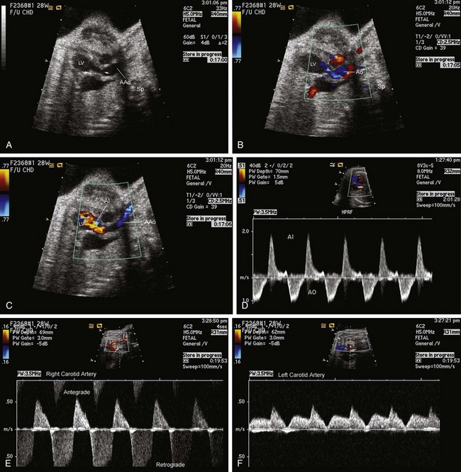
The fetal findings of absent aortic valve leaflets were first described in 1984 upon identification of pandiastolic reversal of flow in the descending aorta.2 Today, direct inspection of the left ventricular outflow tract and ascending aorta will reveal absence of the thin normal leaflets of the aortic valve. The ascending aorta may be dilated as it receives an increased stroke volume with each beat. Prominent pulsations of the ascending and descending aorta are typical clues to the presence of this anomaly.
Postnatal Physiology
Fetal demise is common, but some fetuses do survive to the neonatal period. Aortic insufficiency can worsen upon birth as overall vascular resistance is increased with removal of the low-resistance placental circulation. Hypotension, acidosis, and hypoxemia are to be expected.3
Postnatal Management
If the fetus survives to term, immediate postnatal surgical intervention in cases of hemodynamically severe aortic insufficiency may be helpful; however, there are no reports of such an undertaking. When absent aortic valve leaflets are seen in association with hypoplasia of the left ventricle, the volume of regurgitation is small and may not result in a substantial steal. A staged reconstructive approach using the Norwood operation leading to a single-ventricle palliation has been reported.8 In this case, the aortic annulus was small and was left untouched during the reconstruction. The child has survived through Fontan operation as well, further suggesting that the degree of insufficiency is not of consequence, likely related to the “protective” hemodynamics of a small, stiff left ventricle.9
Outcomes
Imaging Essentials and Important Points
1 Toews WH, Lortscher RH, Kelminson LL. Double outlet right ventricle with absent aortic valve. Chest. 1975;68:381-382.
2 Bierman FZ, Yeh MN, Swersky S, Martin E, Wigger JH, Fox H. Absence of the aortic valve: antenatal and postnatal two-dimensional and Doppler echocardiographic features. J Am Coll Cardiol. 1984;3:833-837.
3 Lin AE, Chin AJ. Absent aortic valve: a complex anomaly. Pediatr Cardiol. 1990;11:195-198.
4 Miyabara S, Ando M, Yoshida K, Saito N, Sugihara H. Absent aortic and pulmonary valves: investigation of three fetal cases with cystic hygroma and review of the literature. Heart Vessels. 1994;9:49-55.
5 Marek J, Skovranek J, Povysilova V. Congenital absence of aortic and pulmonary valve in a fetus with severe heart failure. Heart. 1996;75:98-100.
6 Hartwig NG, Vermeij-Keers C, De Vries HE, Gittenberger-De Groot AC. Aplasia of semilunar valve leaflets: two case reports and developmental aspects. Pediatr Cardiol. 1991;12:114-117.
7 Eronen M, Heikkila P. Absent aortic and dysplastic pulmonary valves associated with ventricular septal defect in fetal hydrops. Pediatr Cardiol. 2003;24:400-402.
8 Harada Y, Takeuchi T, Satomi G, Yasukouchi S. Absent aortic valve: successful palliation in the neonate. Ann Thorac Surg. 1998;66:935-936.
9 Hibino N, Harada Y, Hiramatsu T, Yasukochi S, Satomi G. Fontan operation for hypoplastic left heart syndrome with absent aortic valve. J Thorac Cardiovasc Surg. 2004;128:315-316.