Cone Dystrophy
Clinical Features:
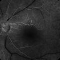
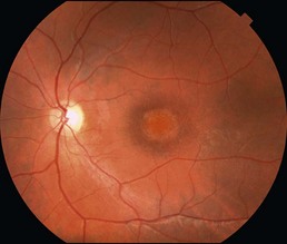
The clinical appearance can vary, but a central bulls eye-type maculopathy is most characteristic (Fig. 16.4.1). Early disease may present with a normal clinical examination. Symptoms include loss of visual acuity, color vision, and hemeralopia.
OCT Features:
There is initially loss of the outer retina and photoreceptors within the central macula (Fig. 16.4.2). Over time, this can progress to complete atrophy. The peripheral macula and retinal periphery appear normal.
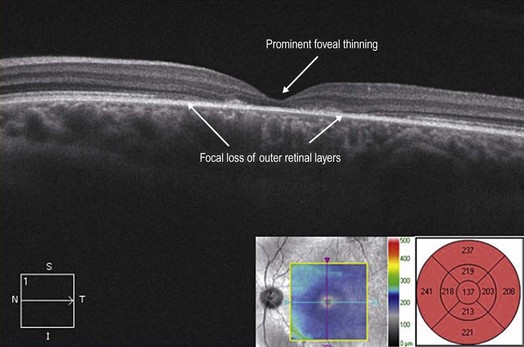
Figure 16.4.2 OCT (corresponding to Figure 16.4.1) shows focal central loss of the outer retinal layers (between arrowheads). There is prominent thinning in the fovea. The corresponding thickness map accentuates the degree of central macular thinning.