14 Computed tomography (CT) scanning
Definition of computed tomography scanning
| Is the process of producing a cross-sectional image of the body by using a collimated beam of radiation that rotates round the patient. Some of the radiation is absorbed and scattered by the body and some is transmitted through the patient and is collected by a number of detectors which are linked to photomultipliers. A signal is then sent to a computer which calculates the amount of radiation absorbed by the patient and reconstructs an image which can then be viewed on a television monitor |
| Dynamic CT | When a number of scans are performed in rapid succession, e.g. to demonstrate blood flow |
| Enhanced CT | The use of a contrast agent to improve the appearance of vessels or organs that are similar in density to the surrounding tissues |
| Field of View | The part of the scanned plane which may be included in the final image |
| Helical | Spiral |
| Image Acquisition | The collection of data in order to produce an image |
| Image Format | The process of storing an image, on computer disk, magnetic tape, film or on the World Wide Web |
| Image Manipulation | To digitally change the appearance of the acquired image in order to improve it |
| Image Reconstruction | The process of generating an image from raw data or a set of unprocessed measurements |
| Isotropic | Having the same properties in all directions, e.g. density |
| Matrix | The columns and rows that form a digital image |
| Mean Window Level | The average range of pixel values in an image |
| Noise | Anything that distracts from the information required on an image |
| Nutating Detector Ring | When the detectors vibrate in such a way as to keep the detectors nearest the tube out of the way of the X-ray beam |
| Pitch | The table movement during one complete rotation of 360° divided by the column width (or slice thickness) |
| Example If the table moves 10 mm during one complete rotation and a beam width of 5 mm was used, the pitch value = 2 |
|
| Pixel | A two dimensional ‘picture cell’ or ‘dot’ that makes up the image on a digital display screen |
| Profile | Line of data |
| Slice | A section through the patient which is recorded when the X-ray tube and detector make one complete rotation |
| Slice Interval | The distance between reconstructed slices |
| Spatial Resolution | The smallest part of an image that can be seen |
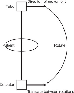
| Translate | Movement in a horizontal direction |
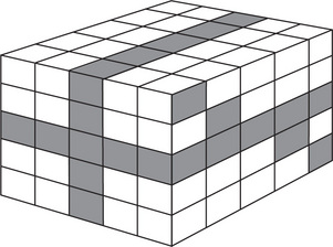
| Voxel | A three dimensional pixel |
| Window | The range of colour (or grey) scale values displayed on a digital image |
| Window Width | The range of pixel values displayed in the digital image |
| Gantry | A circular device for holding the detectors |
| X-ray Tube | A method of producing X-rays which are collimated so that they are aligned to a specific number of detectors |
| Detectors | |
| Photomultipliers | |
| Photodiode | |
| Housing | |
| Movement | |
| Table | For the patient to lie on and can move forward at a predetermined distance or at a constant speed to enable the next ‘slice’ to be taken |
| Operator Console | Where the operator can determine the settings for the scan |
| Display Station | For the viewing, analysis, networking and storage of the final image |
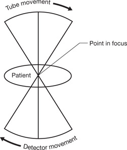
| Tomographic Principle | |
| Tissue Attenuation | As different parts of the body have different densities they absorb radiation to different degrees |
| Attenuation Coefficient | |
| CT Number | Calculated as:
|
| Hounsfield Unit (HU) | |
| Voxel Display | |
| Window | |
| Windowing | |
| Narrow Window | |
| Wide Window | |
| Scanning | |
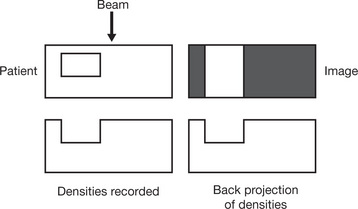
| Detector Action | |
| Image Reconstruction | Diagram A |
| Diagram B | |
| Diagram C | |
| If this process is repeated many hundreds of times, a cross-section through the body can be produced. A minimum display would be a matrix of 512 × 512 = 262 144 pixels | |
| Multiplanar Reformatted Imaging |


| Segmentation | |
| Rendering | |
| Surface rendering | |
| Volume rendering |
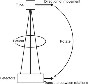
| First Generation Scanner | |
| Second Generation Scanner | |
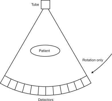
| Third Generation Scanner | |
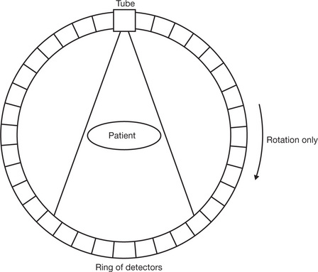
| Fourth Generation Scanner | |
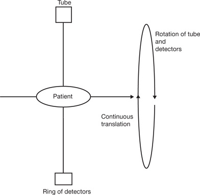
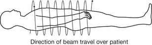
| Helical Scanner |
• The table and patient move slowly and evenly into the gantry until area of interest has been scanned
|
| Multi-slice Scanners |
• Central ray of the X-ray beam follows a spiral round the patient’s body at a speed of 120 rotations per minute
• The tube and several rows of up to 64 detectors (arranged in the direction of the table travel) rotate together
|
| Multi-detector-row CT | |
| Dual Source CT | |
| Fluoroscopic CT |
| Connor S E J, Guest P 1999 Spiral computed tomography pulmonary angiography. Synergy, April | |
| Harvey D 2004 CT evolution. Radiology Today 5(18):August | |
| Hogg P 2005 An overview of PET/CT and its place in today’s UK healthcare system. Synergy, December | |
| Patel H, Clarkson L M 2006 MDCT: Practical implementation of a new technology. Synergy, March | |
| Robinson L 1999 Evaluating the colon with CT. Synergy, August | |
| Scally A, Webster M 2001 The evolution of CT technology and its applications. Synergy, March | |
| Smyth J, Hutchinson J 2004 Cardiac CT: A radiographer’s perspective. Synergy, October |
