CHAPTER 18 Complications of Elbow Arthroscopy
Arthroscopic surgery is increasingly used in the treatment of elbow pathology, including loose bodies, synovitis, degenerative joint disease, osteochondritis dissecans, arthrofibrosis, lateral epicondylitis, olecranon bursitis, fractures, and plica.1,2 Advantages of elbow arthroscopy over open surgery include decreased scarring, decreased risk of infection, less postoperative pain, and better visualization of the joint.2 Elbow arthroscopy is considered one of the most challenging types of arthroscopic surgery, most likely because of the high congruence of the joint and the proximity of neurovascular structures.2
Little research has been done in regard to complications in elbow arthroscopy. Most studies in the literature are case reports or case series that do not specifically look at the incidence of complications of arthroscopic surgery of the elbow. Few large series studies have looked at complications in elbow arthroscopy. In a review of the literature, Savoie3 found 16 reported complications in 465 elbow arthroscopic surgeries, or a 3% incidence. Micheli and colleagues4 described elbow arthroscopy performed on an athletically active pediatric population and found no complications for 47 patients. The members of the Arthroscopy Association of North America were surveyed about the complications of arthroscopic surgery. For the 1569 elbow arthroscopies in the survey, only three complications were reported.5
Kelly and coworkers2 looked at 473 elbow arthroscopies over an 18-year period done by 12 different surgeons and reported an 11% minor complication rate and a 0.8% major complication rate. Savoie and associates6 found an overall complication rate of 7% in a series of 269 consecutive elbow arthroscopies over a 3-year period. In this chapter, we discuss the complications associated with elbow arthroscopy, their possible causes, and tips on how to prevent them.
COMPLICATIONS
The anatomy of the elbow makes arthroscopy a technically difficult procedure that is predisposed to complications.7 The reported complications of elbow arthroscopy include infection, heterotopic ossification, complex regional pain syndrome, nerve injury, fistula, and olecranon bursitis.2,6–10
Infection
One of the most common complications of elbow arthroscopy is infection. The anatomy of the elbow makes it more vulnerable than other joints to infection. The soft tissue envelope around the elbow is extremely thin, and the capsule is separated from the skin by a thin layer of subcutaneous tissue, predisposing the site to prolonged drainage,7 which may precede cellulitis, abscesses, intraarticular infections, or portal fistulas.
Superficial infections and persistent drainage after elbow arthroscopy are much more common than deep infections.2,10,11 Superficial infections of the elbow after arthroscopy typically manifest as prolonged serous drainage or erythema around a portal site. Patients may have low-grade fevers and tenderness around portal sites. The erythema and drainage usually resolve with 2 weeks of oral antibiotics. Immobilization may be beneficial in the setting of prolonged drainage.
Kelly and colleagues2 reported a 5% incidence of prolonged drainage from portal sites and a 2% incidence of superficial infection in their series of 473 procedures. These minor infections resolved with a short course of oral antibiotics. Reddy and associates12 reported a 1% incidence of persistent drainage from arthroscopic portals. Drainage was treated successfully with 7 days of oral antibiotics. Thomas and coworkers6 found a 2.2% incidence of superficial infections, which resolved within 7 to 14 days with oral antibiotics. Several reports2,6,7 indicate that the lateral portals, including the soft spot portal and the anterolateral portal, are more susceptible to infection and prolonged drainage than the medial portals. This finding is probably reflects the fact that the skin is thinner on the lateral side of the elbow.2,7 Suture closure of the portal sites has been recommended to decrease the incidence of prolonged drainage and infection.2,13
Deep infections are rare after elbow arthroscopy. Micheli and colleagues,4 Reddy and associates,12 and Lynch and coworkers14 reported no deep infections in their series of 47, 187, and 21 respective elbow arthroscopies. Thomas and colleagues6 reported one deep infection in their series of 269 patients (0.4% incidence) that resolved with arthroscopic irrigation and débridement, drain placement, and 6 weeks of intravenous antibiotics. Kelly and associates2 reported a 0.8% incidence of intra-articular infections after elbow arthroscopy. They theorized that immediate postoperative steroid injections might increase the risk of joint infections.
Portal fistula formation, although rare, has been reported in the literature. Thomas and colleagues5 reported one soft spot portal fistula formation in a paraplegic patient with a prior instance of methicillin-resistant Staphylococcus aureus infection. The patient was treated successfully with open irrigation, débridement, fistula excision, and closure. The patient also received a course of oral antibiotics.
Nerve Injury
Of all the complications resulting from elbow arthroscopy, nerve injuries are the most feared and the most reported. Most reported nerve injuries associated with elbow arthroscopy are transient, although permanent nerve injuries have been reported.11
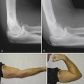
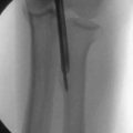
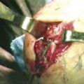
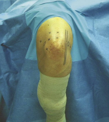
Knowledge of the anatomy of the elbow is crucial to the avoidance of neurologic injury in elbow arthroscopy. All three nerves that cross the elbow joint are close to the capsule, and capsular distention does not protect the nerves from capsular procedures, such as a release or synovectomy.7 Lynch and coworkers14 performed cadaveric dissections and found that the radial nerve lay within an average of 5 mm of the joint and that distention of the joint increased this distance to 10 mm. The median nerve was also found to be within 5 mm of the anterior joint. Lindenfeld20 reported that the radial nerve was an average of 7.8 mm from the anterolateral portal when the portal was made 3 cm distal and 1 cm anterior to the lateral epicondyle (Fig. 18-1). The radial nerve and the posterior interosseous nerve are therefore at risk when making an anterolateral portal. The ulnar nerve is at risk when creating the anteromedial portal. The median nerve is at risk when débriding or performing a capsulectomy of the anterior capsule.7 O’Driscoll and Morrey10 reported three patients with transient radial nerve palsies lasting several hours in their series of 71 elbow arthroscopies. They attributed these palsies to local anesthetic extravasation. Lynch and associates14 reported a 14% incidence of nerve injuries in their series of 21 arthroscopies, including a transient radial nerve palsy, a transient median nerve palsy, and a neuroma of the medial antebrachial cutaneous nerve.
Kelly and colleagues2 had a 2% incidence of transient nerve palsies in their series of 473 consecutive elbow arthroscopies. These palsies included four superficial radial, five ulnar, one posterior interosseous, one anterior interosseous, and one medial antebrachial cutaneous nerve palsies. All palsies resolved within 6 weeks after arthroscopy, except for one case, which resolved within 6 months. They reported that rheumatoid arthritis and capsular release were risk factors for nerve injury. Thomas and coworkers6 reported a 1.9% incidence of transient nerve palsies in a series of 269 arthroscopies and identified an increased risk of nerve injury with capsular release. The nerve palsies in this series required an average of 6.1 months to completely resolve.
Permanent nerve injuries after elbow arthroscopy are a rare complication. Neither series by Kelly and colleagues2 or Thomas and associates6 reported a permanent nerve injury. Reddy and coworkers12 reported a complete transection of the ulnar nerve after an arthroscopic synovectomy and loose body removal. The injury was not noticed during the initial procedure, but after re-exploration of the ulnar nerve 3 days postoperatively, a 1.5-cm defect in the ulnar nerve was found and repaired. The patient regained some light touch and pinprick sensation but did not regain strength in the intrinsic musculature. Casscells16 reported an irreparable injury to the ulnar nerve when using a motorized shaver posteromedially. Jones and Savoie,17 Papilion and colleagues,18 and Thomas and associates19 reported permanent posterior interosseous nerve injuries that occurred during arthroscopic procedures. Jones and Savoie17 described the injury in a patient who had a displaced radial head fracture that healed to the anterior capsule. During arthroscopic débridement and manipulation, the capsule separated in this area rather than near the humerus, where it had been excised, and it severed the nerve. Jones and Savoie17 recommended avoiding arthroscopic capsular release in patients with excessive scarring in the vicinity of the posterior interosseous nerve without first dissecting out the nerve.
To avoid nerve injuries in elbow arthroscopy, several principles should be followed. Joint distention should be used before portal placement to move the nerves farther away from the joint. Even so, the nerves remain close to the capsule, and distention does not protect the nerves during capsular procedures. Proximal portals are safer than more distal portals, and the radial nerve is most at risk when making the anterolateral portal. The ulnar nerve should be palpated before anteromedial portal placement. Posteromedial and direct anterior portals should never be used. The elbow should be flexed to 90 degrees during portal placement. Retractors may increase the safety of elbow arthroscopic procedures, especially in capsulectomies and synovectomies. Particular caution should be used when working posteromedially because of the proximity of the ulnar nerve. Suction should be avoided around nerves, and instrument tips should be visualized at all times. Knowledge of elbow anatomy and proficiency in arthroscopy are keys to avoiding neurologic injury in elbow arthroscopy.7,15,20
Other Complications
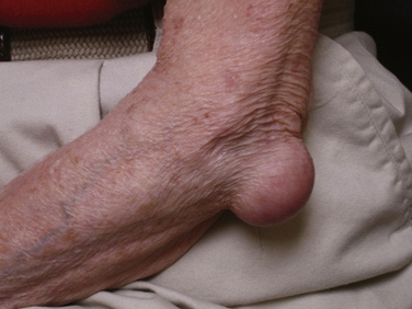
A 1.1% incidence of olecranon bursitis was reported by Thomas and colleagues6 in their series. Most cases were benign and responded to conservative therapy (Fig. 18-2). No other incidents of olecranon bursitis associated with elbow arthroscopy have been reported in the literature.
Kelly and associates2 reported seven cases of minor postoperative contracture in their series of 473 elbow arthroscopies. The patients lost less than 20 degrees, and the contractures only affected the flexion-extension plane. No heterotopic ossification occurred. Thomas and colleagues6 reported one case of anterior heterotopic ossification after an arthroscopic ulnohumeral arthroplasty and capsular release. Less than 20 degrees of motion was lost due to the heterotopic ossification.
Complex regional pain syndrome is another rare complication that has been reported after elbow arthroscopy.6 These cases have responded to physical therapy, pain management, and sympathetic blocks.
CONCLUSIONS
Elbow arthroscopy is a safe procedure when performed by experienced surgeons. Major complications, including permanent nerve injuries, are rare, and the incidence of minor complications is comparable to that for other forms of arthroscopy.
1. Baker CL, Jones GL. Arthroscopy of the elbow. Am J Sports Med. 1999;27:251-264.
2. Kelly EW, Morrey BF, O’Driscoll SW. Complications of elbow arthroscopy. J Bone Joint Surg Am. 2001;83:25-34.
3. Savoie FH. Complications. In: Savoie FH, Field LD, editors. Arthroscopy of the Elbow. New York, NY: Churchill Livingstone; 1996:151-156.
4. Micheli LJ, Luke AC, Mintzer CM, Waters PM. Elbow arthroscopy in the pediatric and adolescent population. Arthroscopy. 2001;17:694-699.
5. Small NC. Complications in arthroscopy: the knee and other joints. Arthroscopy. 1986;2:253-258.
6. Thomas RJ, Savoie FH, Field LD. Complications in elbow arthroscopy. Am J Sports Med. 2010. In press
7. Morrey BF. Complications of elbow arthroscopy. Instr Course Lect. 2000;49:255-258.
8. Angelo RL. Advances in elbow arthroscopy. Orthopedics. 1993;16:1037-1046.
9. Redden JF, Stanley D. Arthroscopic fenestration of the olecranon fossa in the treatment of osteoarthritis of the elbow. Arthroscopy. 1993;9:14-16.
10. O’Driscoll SW, Morrey BF. Arthroscopy of the elbow: diagnostic and therapeutic benefits and hazards. J Bone Joint Surg Am. 1992;74:84-94.
11. Phillips BB. Arthroscopy of the upper extremity. In: Canale ST, Beaty JH, editors. Campbell’s Operative Orthopaedics. Philadelphia, PA: Mosby Elsevier; 2008:2923-3014.
12. Reddy AS, Kvitne RS, Yocum LA, et al. Arthroscopy of the elbow: a long term clinical review. Arthroscopy. 2000;16:588-594.
13. Morrey BF, Askew LJ, An KN, et al. A biomechanical study of normal functional elbow motion. J Bone Joint Surg Am. 1981;63:872-877.
14. Lynch GJ, Meyers JF, Whipple TL, et al. Neurovascular anatomy and elbow arthroscopy: inherent risks. Arthroscopy. 1986;2:190-197.
15. Lindenfeld TN. Medial approach in elbow arthroscopy. Am J Sports Med. 1990;18:413-417.
16. Casscells SW. Neurovascular anatomy and elbow arthroscopy: inherent risks [editor’s comment]. Arthroscopy. 1987;2:190.
17. Jones GS, Savoie FHIII. Arthroscopic capsular release of flexion contractures (arthrofibrosis) of the elbow. Arthroscopy. 1993;9:277-283.
18. Papilion JD, Neff RS, Shall LM. Compression neuropathy of the radial nerve as a complication of elbow arthroscopy: a case report and review of the literature. Arthroscopy. 1988;4:284-286.
19. Thomas MA, Fast A, Shapiro DL. Radial nerve damage as a complication of elbow arthroscopy. Clin Orthop. 1987;215:130-131.
20. Wiesler ER, Poehling GG. Elbow arthroscopy: introduction, indications, complications, and results. In: McGinty JB, ed. Operative Arthroscopy. Philadelphia, PA: Lippincott Williams & Wilkins; 2003:661-664.