Chapter 307 Common Lesions of the Oral Soft Tissues
Oropharyngeal Candidiasis
Oropharyngeal infection with Candida albicans (thrush, moniliasis) (Chapter 226.1) is common in neonates from contact with the organism in the birth canal or breast. The lesions of oropharyngeal candidiasis (OPC) appears as white plaques covering all or part of the oropharyngeal mucosa. These plaques are removable from the underlying surface, which is characteristically inflamed and has pinpoint hemorrhages. The diagnosis is confirmed by direct microscopic examination on potassium hydroxide smears and culture of scrapings from lesions. OPC is usually self-limited in the healthy newborn infant, but topical application of nystatin to the oral cavity of the baby and to the nipples of breast-feeding mothers will hasten recovery.
Aphthous Ulcers
The aphthous ulcer (canker sore) is a distinct oral lesion, prone to recurrence. The differential diagnosis is noted in Table 307-1. Aphthous ulcers are reported to develop in 20% of the population. Their etiology is unclear, but allergic or immunologic reactions, emotional stress, genetics, and injury to the soft tissues in the mouth have been implicated. Aphthous-like lesions may be associated with inflammatory bowel disease, Behçet disease, gluten-sensitive enteropathy, periodic fever-aphthae-pharyngitis-adenitis syndrome, Sweet syndrome, HIV infection (especially if ulcers are large and slow to heal), and cyclic neutropenia. Clinically, these ulcers are characterized by well-circumscribed, ulcerative lesions with a white necrotic base surrounded by a red halo. The lesions last 10-14 days and heal without scarring. Over-the-counter palliative therapies, such as benzocaine and topical lidocaine, are effective, as well as topical steroids. Tetracycline has been shown to have benefit with severe outbreaks, but caution is necessary in pregnant women and young children to prevent tetracycline tooth staining during a child’s tooth development.
| CONDITION | COMMENT |
|---|---|
| COMMON | |
| Aphthous (canker sore) | Painful, circumscribed lesions; recurrences |
| Traumatic | Accidents, chronic cheek biter, or after dental local anesthesia |
| Hand, foot, mouth disease | Painful; lesions on tongue, anterior oral cavity, hands, and feet |
| Herpangina | Painful; lesions confined to soft palate and oropharynx |
| Herpetic gingivostomatitis | Vesicles on mucocutaneous borders; painful, febrile |
| Recurrent herpes labialis | Vesicles on lips; painful |
| Chemical burns | Alkali, acid, aspirin; painful |
| Heat burns | Hot food, electrical |
| UNCOMMON | |
| Neutrophil defects | Agranulocytosis, leukemia, cyclic neutropenia; painful |
| Systemic lupus erythematosus | Recurrent, may be painless |
| Behçet’s syndrome | Resembles aphthous lesions; associated with genital ulcers, uveitis |
| Necrotizing ulcerative gingivostomatitis | Vincent stomatitis; painful |
| Syphilis | Chancre or gumma; painless |
| Oral Crohn disease | Aphthous-like; painful |
| Histoplasmosis | Lingual |
Herpetic Gingivostomatitis
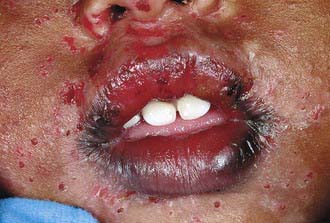
After an initial incubation period of ∼1 wk, the initial infection with herpes simplex virus manifests as fever and malaise, usually in a child <5 yr (Chapter 244). The oral cavity can show various expressions, including the gingiva becoming erythematous, mucosal hemorrhages, and clusters of small vesicles erupting throughout the mouth. There is often involvement of the mucocutaneous margin and perioral skin (Fig. 307-1). The oral symptoms generally are accompanied by fever, lymphadenopathy, and difficulty eating and drinking. The symptoms usually regress within 2 wk without scarring. Fluids should be encouraged because the child may become dehydrated. Analgesics and anesthetic rinses can make the child more comfortable. Oral acyclovir if taken within the first 3 days of symptoms may be beneficial in shortening the duration of symptoms. Caution should be exercised to prevent autoinoculation or transmission of infection to the eyes.
Geographic Tongue
Geographic tongue (migratory glossitis) is a benign and asymptomatic lesion and is characterized by one or more smooth, bright red patches, often showing a yellow, gray, or white membranous margin on the dorsum of an otherwise normally roughened tongue. The condition has no known cause, and no treatment is indicated (Chapter 656).


