[level-membership-for-internal-medicine-category]
Coma
Coma results from sustained impairment of awareness of self and of the environment. It can simply be defined as a state of unconsciousness or numerically categorised as a GCS score of<8.
Causes
Trauma
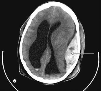
Figure 11 CT of the head.
There is a left-sided extradural haematoma (arrow). The haematoma is confined giving rise to its characteristic biconvex shape, with a well-defined margin.
Metabolic
Organ Failure
Infective
Toxin/Drug-Induced
History
The history for the comatose patient is usually obtained co-laterally from relatives, friends, witnesses or ambulance crew. Other sources of information are previous hospital notes, records kept by general practitioners and tablets or prescriptions that have been found at the patient’s premises.
Presentation
The circumstances regarding discovery of the patient is usually the first piece of information to be reported. Trauma patients may have been transported from sites of road traffic accidents, fire or have been found assaulted on the street. When teenagers are brought in unconscious from a night club, it is important to exclude alcohol intoxication, epilepsy, hypoglycaemia and the effects of illicit drugs (e.g. malignant hyperthermia with ‘ecstasy’). Attempted suicides may have an accompanying note or drug bottles may have been recovered from the scene. Carbon monoxide poisoning occurs with suicide attempts in enclosed areas with running engines or may be a complication in victims involved with fires. Patients who have been brought in from cold environments may also have hypothermia in addition to the primary event.
Onset
When witnessed, information regarding the speed of onset of coma may help in determining a cause. Sudden onset of unconsciousness is characteristic of a seizure or a vascular event.
Trauma
Head injuries are a common cause of coma, but not all causes are due to blunt trauma to the cranium. Diffuse axonal injury results from severe shearing forces on the brain, a sequel to rapid acceleration and deceleration forces. Blunt head injuries may cause extradural haemorrhages as a result of skull fractures with laceration of meningeal arteries. There is usually a history of an injury with transient loss of consciousness, a lucid interval in which the patient feels and appears well, followed by drowsiness, headache, vomiting, progressive hemiplegia and eventually coma. Subdural haemorrhages are a consequence of severe trauma, with cortical lacerations or less severe trauma, with laceration of bridging veins. Chronic subdural haemorrhages may result even in the absence of trauma; elderly patients with cortical atrophy are predisposed to this type of injury.
In addition to head injury, coma may also complicate any other condition severe enough to result in circulatory or respiratory insufficiency.
Headache
The onset of severe headache prior to coma may be caused by trauma, subarachnoid haemorrhage (classically patients complain of a sudden onset of blinding headache, the worst ever experienced) or meningitis (headache associated with photophobia and neck stiffness). Progressive headache, worse in the mornings and associated with vomiting, may be due to raised intracranial pressure from a cerebral tumour.
Predisposing factors
The history of possible predisposing factors is useful when assessing the comatose patient. The presence of diabetes should lead to the consideration of ketoacidosis (type I diabetics) and hypoglycaemia (oral hypoglycaemic or insulin dosage errors). Patients with known hepatic or renal failure may deteriorate to coma as a result of encephalopathy or uraemia, respectively. Coma may also complicate severe hypothyroidism. Previous suicide attempts or a history of depression should lead to the consideration of drug overdose. Epileptic patients may be in status epilepticus or in a post-ictal recovery state. Pre-existing cardiac or respiratory disease may result in coma as a terminal event.
Examination
Temperature
Body temperature should be recorded and a series of normal body temperatures will exclude hypothermia and hyperpyrexia as a consequence of heat stroke or illicit drug use.
General examination
A thorough inspection should be performed. This will necessitate removal of clothing, log rolling (to examine the back) and a detailed examination of the scalp for bleeding, haematomas and fractures. The presence of bilateral periorbital haematomas or cerebrospinal fluid rhinorrhoea may indicate an anterior fossa fracture of the skull. Bruising over the mastoid (Battle’s sign) or cerebrospinal fluid otorrhoea may be due to a middle cranial fossa fracture. Patients with carbon monoxide poisoning may appear bright red. Patients with myxoedema are characteristically obese, with coarse features, dry skin and brittle hair. The arms should be carefully scrutinised for lacerations and needle puncture sites suggestive of drug abuse and thus the possibility of opiate overdose. A petechial rash may be visible with meningococcal meningitis.
The organ systems should be examined in turn; suspect encephalopathy if signs suggestive of liver disease are present (p. 37) or uraemia with chronic renal failure. The pulse should be examined for arrhythmias, which may precipitate cardiac failure. The JVP may also be elevated. Other causes of JVP distension include tension pneumothorax and cardiac tamponade, both of which may embarrass the circulation and precipitate unconsciousness.
Auscultation of the chest may reveal bilateral crepitations with pulmonary oedema from left ventricular failure, bronchopneumonia, chronic bronchitis or aspiration.
Neurological examination
The neurological examination should begin with the GCS score. Patients who are not involved in trauma should be assessed for neck stiffness which may result from meningitis or subarachnoid haemorrhage. Trauma patients should have the cervical spine assessed for injury before any manipulation is undertaken. The pupils should be examined for size and light reflex. Pinpoint pupils occur with opiate overdoses, small pupils with brainstem lesions and dilated pupils with cocaine or amphetamine use, hypoglycaemia, post-ictal states and brainstem death. A unilateral fixed and dilated pupil occurs with an oculomotor nerve lesion, which may result from pressure exerted from intracranial haemorrhage or tumour. Fundoscopy should be performed to identify loss of retinal vein pulsation or frank papilloedema indicating raised intracranial pressure. A limited neurological examination can be performed on comatose patients; this will include assessment of tone, reflexes and the Babinski response. Unilateral increased tone, hyperreflexia and an up-going plantar response is indicative of a contralateral upper motor neurone lesion, such as a stroke, intracranial haemorrhage or tumour.
General Investigations
■ FBC
↑ WCC with meningitis. Viral meningitis is associated with lymphocytosis, and neutrophilia is associated with bacterial meningitis.
■ Blood glucose
A bedside blood sugar estimation is very useful to exclude hypoglycaemia.
■ Urinalysis
Ketones will be present in diabetic ketoacidosis. The urine should also be sent for toxicology analysis if poisoning is suspected.
■ U&Es
The urea and creatinine is elevated with renal failure. Disorders of sodium levels will be readily identified.
■ LFTs
The bilirubin and transaminases are markedly elevated in liver failure.
■ TFTs
↑ TSH, ↓ T4 with hypothyroidism.
■ Toxicology screen
For cases of suspected poisoning and overdose, serum levels of salicylates and paracetamol will be helpful in both diagnosis and treatment. Ethanol levels should also be checked, and carboxyhaemoglobin levels if carbon monoxide poisoning is suspected.
■ ECG
An ECG is useful to diagnose arrhythmia and myocardial infarction (p. 62); either disorder may compromise the circulation.
■ CXR
May reveal pulmonary oedema or lobar consolidation due to infection or aspiration.
■ X-ray cervical spine
Exclude fracture in traumatic causes of coma.
■ CT
With a history of trauma or clinical signs which suggest head injury, a CT scan should be requested (this is in preference to a skull X-ray). Additionally, CT will reveal the presence and location of intracranial bleeding and tumours.
Specific Investigations
■ Lumbar puncture
A lumbar puncture is performed in cases of suspected meningitis and the CSF (which may be turbid) is then sent for microscopy and culture.
■ EEG
The EEG is indicated when the diagnosis of epilepsy is suspected. Status epilepticus, however, should be diagnosed and treated clinically. The EEG is also useful in the diagnosis of encephalitis.
[/level-membership-for-internal-medicine-category][not-level-membership-for-internal-medicine-category]
Coma
Coma results from sustained impairment of awareness of self and of the environment. It can simply be defined as a state of unconsciousness or numerically categorised as a GCS score of<8.
Causes
Trauma
Figure 11 CT of the head.
There is a left-sided extradural haematoma (arrow). The haematoma is confined giving rise to its characteristic biconvex shape, with a well-defined margin.
Metabolic
Organ Failure
Infective
Toxin/Drug-Induced
History
The history for the comatose patient is usually obtained co-laterally from relatives, friends, witnesses or ambulance crew. Other sources of information are previous hospital notes, records kept by general practitioners and tablets or prescriptions that have been found at the patient’s premises.
Presentation
The circumstances regarding discovery of the patient is usually the first piece of information to be reported. Trauma patients may have been transported from sites of road traffic accidents, fire or have been found assaulted on the street. When teenagers are brought in unconscious from a night club, it is important to exclude alcohol intoxication, epilepsy, hypoglycaemia and the effects of illicit drugs (e.g. malignant hyperthermia with ‘ecstasy’). Attempted suicides may have an accompanying note or drug bottles may have been recovered from the scene. Carbon monoxide poisoning occurs with suicide attempts in enclosed areas with running engines or may be a complication in victims involved with fires. Patients who have been brought in from cold environments may also have hypothermia in addition to the primary event.
[/not-level-membership-for-internal-medicine-category]
