Colposcopy

Published on 31/05/2015 by admin

Filed under Obstetrics & Gynecology

Last modified 22/04/2025

Print this page

rate 1 star rate 2 star rate 3 star rate 4 star rate 5 star
Your rating: none, Average: 3 (1 votes)

This article have been viewed 2168 times

Chapter 238 Colposcopy

TECHNIQUE

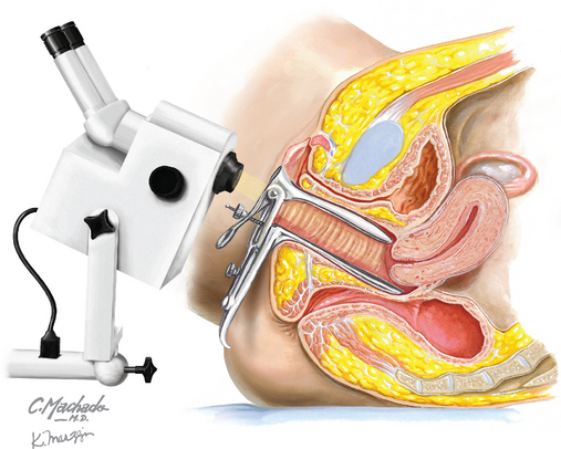
The patient is placed in the dorsal lithotomy position. (If a bimanual examination is performed, the amount of lubricant used should be limited because this lubricant, and glove powder, can adversely affect cytologic studies.) By use of the largest warmed but unlubricated speculum the patient can comfortably accommodate, the cervix should be brought into full view. Following gross inspection for lesions, excessive secretions may be gently blotted away, cultures obtained, or a repeat Papanicolaou (Pap) smear taken.

The colposcope should be positioned to provide an unobstructed view of the cervix and maintained in a position and height that is comfortable for both the patient and the examiner. Acetic acid (3% to 5%) should be liberally applied to the cervix with large cotton-tipped applicators, cotton balls, or small gauze sponges. Acetic acid causes the columnar epithelial cells to swell and opacifies metaplastic and dysplastic cells. The changes brought on by the application of acetic acid are only temporary, requiring periodic reapplication at roughly 5-minute intervals.

Inspection of the cervix begins using the lowest magnification, with additional magnification added later, if needed. The transformation zone should be identified and inspected in its entirety. If necessary, the cervix may be manipulated using an acetic acid–soaked applicator stick, a cervical hook (similar to a skin hook retractor), or an endocervical speculum.

For a colposcopy to be considered “adequate” the entire transformation zone must be visualized. The full extent of any lesion present must also be visible for the study to be considered adequate. If the colposcopy is “inadequate,” diagnostic conization will be required. Any areas of white change, vascular abnormality, or mosaicism should be inspected under greater magnification. Vascular patterns may be enhanced by the interposition of a green filter in the colposcope’s light path, making the vessels appear black against the pale background of the epithelium.

Any area of abnormality identified should be biopsied. (Although rarely necessary, abnormal areas may be stained with Lugol solution to aid this identification.) When multiple abnormalities are present, biopsies of the most severe areas take precedence. Whenever possible, the biopsy should include the edge or border of the lesion. Biopsies should be placed in a buffered formalin solution for transport to the pathology laboratory.

Curettage of the endocervical canal (ECC) should generally be included to exclude the possibility of endocervical lesions above the limits of visibility. The ECC is especially helpful as a first stage in the evaluation of atypical glandular cells.

If bleeding from a biopsy site persists or is heavy, Monsel solution may be applied. Monsel solution should be applied only after all specimens have been obtained.

For colposcopy of the vulva, a weaker concentration of acetic acid will result in less burning and discomfort. Because of the relatively thicker epithelium of the vulva, the acetic acid must be left in contact with the tissues for a longer period (even if the stronger solution is chosen). Soaking a gauze sponge and allowing it to remain in contact with the skin for several minutes most easily accomplish this.

REFERENCES

Level III

American College of Obstetricians and Gynecologists. Diagnosis and treatment of cervical carcinomas. ACOG Practice Bulletin 35. Obstet Gynecol. 2002;99:855.

American College of Obstetricians and Gynecologists. Cervical cytology screening. ACOG Practice Bulletin 45. Obstet Gynecol. 2003;102:417.

American College of Obstetricians and Gynecologists. Cervical cancer screening in adolescents. ACOG Committee Opinion 300. Obstet Gynecol. 2004;104:885.

American College of Obstetricians and Gynecologists. Human papillomavirus. ACOG Practice Bulletin 61. Obstet Gynecol. 2005;105:905.

American College of Obstetricians and Gynecologists. Management of abnormal cervical cytology and histology. ACOG Practice Bulletin 66. Obstet Gynecol. 2005;106:645.

American College of Obstetricians and Gynecologists. Evaluation and management of abnormal cervical cytology and histology in the adolescent. ACOG Committee Opinion 330. Obstet Gynecol. 2006;107:963.

Burke L, Antonioli DA, Ducatman BS. Colposcopy: Text and atlas. East Norwalk, Conn: Appleton & Lange, 1991.

Craine BL, Craine ER. Digital imaging colposcopy: basic concepts and applications. Obstet Gynecol. 1993;82:869.

Davis GD. Colposcopic examination of the vagina. Obstet Gynecol Clin North Am. 1993;20:217.

Lau S, Franco EL. Management of low-grade cervical lesions in young women. CMAJ. 2005;173:771.

Rickert VI, Kozlowski KJ, Warren AM, et al. Adolescents and colposcopy: the use of different procedures to reduce anxiety. Am J Obstet Gynecol. 1994;170:504.

Safaeian M, Solomon D, Wacholder S, et al. Risk of precancer and follow-up management strategies for women with human papillomavirus-negative atypical squamous cells of undetermined significance. Obstet Gynecol. 2007;109:1325.

Shafi MI, Dunn JA, Chenoy R, et al. Digital imaging colposcopy, image analysis and quantification of the colposcopic image. Br J Obstet Gynaecol. 1994;101:234.

Wright TCJr, Cox JT, Massad LS, et al. 2001 Consensus Guidelines for the management of women with cervical cytological abnormalities. JAMA. 2002;287:2120.

Wright VC. Understanding the colposcope. Optics, light path, magnification, and field of view. Obstet Gynecol Clin North Am. 1993;20:31.