[level-membership-for-basic-science-category]
20 Cochlear nerve
Auditory System
The cochlea
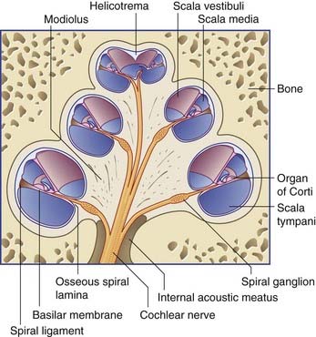
The main features of cochlear structure are seen in Figures 20.1 and 20.2. The cochlea is pictured as though it were upright, but in life it lies on its side, as shown earlier in Figure 19.1. The central bony pillar of the cochlea (the modiolus) is in the axis of the internal acoustic meatus. Projecting from the modiolus, like the flange of a screw, is the osseous spiral lamina. The basilar membrane is attached to the tip of this lamina; it reaches across the cavity of the bony cochlea to become attached to the spiral ligament on the outer wall. The osseous spiral lamina and spiral ligament become progressively smaller as one ascends the two and one half turns of the cochlea, and the fibers of the basilar membrane become progressively longer.
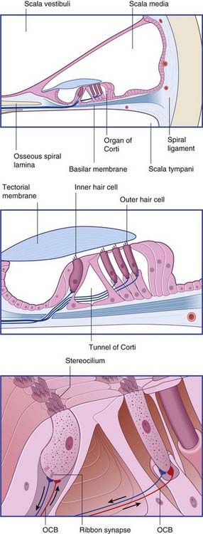
The outer hair cells are contractile (in tissue culture), and they have substantial efferent nerve endings (Figure 20.2). In theory, at least, oscillatory movements of outer hair cells could influence the sensitivity of the inner hair cells through effects on the tectorial or basilar membrane.
Sound transduction
The vibrations of the tympanic membrane in response to sound waves are transmitted along the ossicular chain. The footplate of the stapes fits snugly into the oval window, and vibrations of the stapes are converted to pressure waves in the scala vestibuli. The pressure waves are transmitted through the vestibular membrane to reach the basilar membrane. High-frequency pressure waves, created by high-pitched sounds, cause the short fibers of the basilar membrane in the basal turn of the cochlea to resonate and absorb their energy. Low-frequency waves produce resonance in the apical turn where the fibers are longest. The basilar membrane is therefore tonotopic in its fiber sequence. Not surprisingly, the inner hair cells have a similar tonotopic sequence. In response to local resonance, the cells become depolarized and liberate excitatory transmitter substance from synaptic ribbons (Figure 20.2).
Central auditory pathways
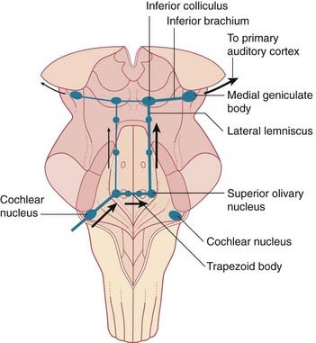
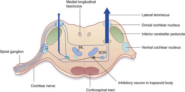
The general plan of the central auditory pathway from the left cochlear nerve to the cerebral cortex is shown in Figure 20.3. The first cell station is the cochlear nuclei, where all cochlear nerve fibers terminate upon entry to the brainstem. From here, some second-order fibers project all the way to the opposite inferior colliculus by way of the trapezoid body and lateral lemniscus. The inferior brachium links the inferior colliculus to the medial geniculate body, which projects to the primary auditory cortex in the temporal lobe.
Functional anatomy (Figure 20.4)
Inferior colliculus
In addition to projecting to the medial geniculate body (nucleus), the inferior colliculus exerts inhibitory effects on its opposite number through the collicular commissure (Figure 20.3). It also contributes to the tectospinal tract.
Medial geniculate body
The medial geniculate body is the specific thalamic nucleus for hearing. The main (ventral) nucleus is laminated and tonotopic, and its large (magnocellular) principal neurons project as the auditory radiation to the primary auditory cortex (Figure 20.5).
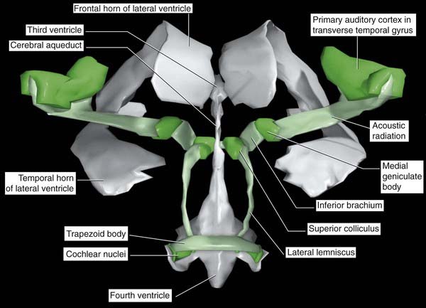
Figure 20.5 Graphic reconstruction of the central auditory pathways from a postmortem brain.
(Reproduced from Kretschmann, H-J. and Weinrich, W. Neurofunctional Systems: 3D Reconstructions with Correlated Neuroimaging: Text and CD-ROM. New York: Thieme 1998, with kind permission of the authors and the publisher.)
Primary auditory cortex
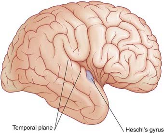
The upper surface of the temporal lobe shows two or more transverse temporal gyri. The anterior one (the gyrus of Heschl) contains the primary auditory cortex (Figure 20.6). Tonotopic arrangement is preserved in Heschl’s gyrus, its posterior part being responsive to high tones and its anterior part to low tones. The auditory cortex responds to auditory stimuli within the contralateral sound field. In cats, destruction of a patch of primary cortex on one side produces a sigoma or ‘deaf spot’ in the contralateral sound field. In humans, ablation of the superior temporal gyrus (in the course of tumor removal) does not cause deafness, but it significantly reduces ability to judge the direction and distance of a source of sound.
Brainstem auditory evoked potentials are described in Chapter 31.
Brainstem acoustic reflexes
Deafness
Deafness is a widespread problem in the community. About 10% of adults suffer from it in some degree. The cause may lie in the outer, middle, or inner ear, or in the cochlear neural pathway. The two fundamental types of deafness are described in Clinical Panel 20.1.
Clinical Panel 20.1 Two kinds of deafness
Ototoxic deafness may follow administration of drugs, including streptomycin, neomycin, and quinine.
An important cause of sensorineural deafness in adults is an acoustic neuroma. Because the trigeminal and facial nerves may be affected as well as the cochlear and vestibular, this tumor is described in Chapter 22.
Cooper NP, Cooper JJ. Efferent-mediated control of basilar membrane movement. J Physiol. 2006;576:49-54.
Corwin JT, Warchol ME. Auditory hair cells: structure, function, development, and regeneration. Ann Rev Neurosci. 1991;14:301-333.
Howarth A, Shone GR. Ageing and the auditory system. Postgrad Med J. 2006:166-171.
Raphael Y, Altschuler RA. Structure and innervation of the cochlea. Brain Res Bull. 2003;60:397-422.
[/level-membership-for-basic-science-category][not-level-membership-for-basic-science-category]
20 Cochlear nerve
Auditory System
The cochlea
The main features of cochlear structure are seen in Figures 20.1 and 20.2. The cochlea is pictured as though it were upright, but in life it lies on its side, as shown earlier in Figure 19.1. The central bony pillar of the cochlea (the modiolus) is in the axis of the internal acoustic meatus. Projecting from the modiolus, like the flange of a screw, is the osseous spiral lamina. The basilar membrane is attached to the tip of this lamina; it reaches across the cavity of the bony cochlea to become attached to the spiral ligament on the outer wall. The osseous spiral lamina and spiral ligament become progressively smaller as one ascends the two and one half turns of the cochlea, and the fibers of the basilar membrane become progressively longer.
The outer hair cells are contractile (in tissue culture), and they have substantial efferent nerve endings (Figure 20.2). In theory, at least, oscillatory movements of outer hair cells could influence the sensitivity of the inner hair cells through effects on the tectorial or basilar membrane.
Sound transduction
The vibrations of the tympanic membrane in response to sound waves are transmitted along the ossicular chain. The footplate of the stapes fits snugly into the oval window, and vibrations of the stapes are converted to pressure waves in the scala vestibuli. The pressure waves are transmitted through the vestibular membrane to reach the basilar membrane. High-frequency pressure waves, created by high-pitched sounds, cause the short fibers of the basilar membrane in the basal turn of the cochlea to resonate and absorb their energy. Low-frequency waves produce resonance in the apical turn where the fibers are longest. The basilar membrane is therefore tonotopic in its fiber sequence. Not surprisingly, the inner hair cells have a similar tonotopic sequence. In response to local resonance, the cells become depolarized and liberate excitatory transmitter substance from synaptic ribbons (Figure 20.2).
