Clubbing
Clubbing is the selective bulbous enlargement of the distal segments of fingers or toes due to proliferation of connective tissues (Fig. 10).
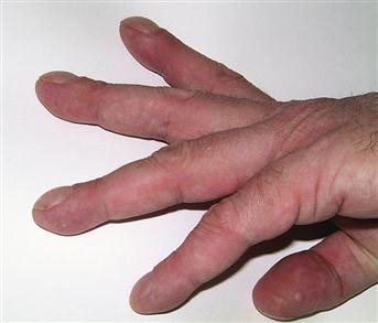
Figure 10 Clubbing.
Note the exaggerated longitudinal curvature and the loss of angle between the nail and the nailbed.
History
A systematic approach to the history is required to determine the cause of clubbing. Respiratory, cardiovascular and gastrointestinal system enquiries should be made in an attempt to determine the underlying cause. A detailed discussion on the diagnosis of congenital cyanotic heart disease (tetralogy of Fallot, transposition of great arteries, total anomalous pulmonary venous drainage) is beyond the scope of this text.
Duration
The duration of clubbing may give an indication of the underlying aetiology. Clubbing present since infancy would suggest a familial trait or congenital cyanotic heart disease. Alternatively, it may be a secondary manifestation of hereditary disorders such as coeliac disease and cystic fibrosis.
Respiratory symptoms
A history of cough, haemoptysis, dyspnoea and weight loss in a chronic smoker should alert the clinician to the diagnosis of bronchial carcinoma. Symptoms of metastasis (bone pain, jaundice) and paraneoplastic involvement (neuropathy, thirst and polyuria from hypercalcaemia) should also be ascertained. With bronchial carcinoma, clubbing may be part of a generalised arthritic manifestation of hypertrophic pulmonary osteoarthropathy.
Cough productive of copious amounts of purulent sputum is usually the main complaint with bronchiectasis. With a chronic lung abscess or empyema, the symptoms are often less specific and may be preceded by a history of pneumonia or aspiration secondary to neurological disorders, dysphagia or alcoholism. Patients may present with a persistent low-grade pyrexia, malaise, weight loss and productive cough. Shortness of breath and a non-productive cough characterise interstitial lung disease.
Gastrointestinal symptoms
Malaise, diarrhoea, abdominal pain and weight loss are experienced by most patients with inflammatory bowel disease and coeliac disease. The presence of aphthous ulceration, fistulae and perianal sepsis are suggestive of Crohn’s disease. Predominantly rectal involvement with tenesmus, mucus and blood PR is more characteristic of ulcerative colitis; however, differentiation based on the history alone can be very difficult. Symptoms of coeliac disease tend to be very variable and non-specific, with non-bloody diarrhoea with steatorrhoea.
Heavy alcohol consumption or previous infection with hepatitis B will predispose to cirrhosis of the liver. Patients may complain of jaundice, abdominal distension due to ascites (p. 35), upper gastrointestinal haemorrhage due to varices or bruising from impaired clotting (p. 548). Severely affected individuals may present with encephalopathy or even coma (p. 69).
Family history
A family history of clubbing or cystic fibrosis will suggest a hereditary cause. Approximately 15% of patients with coeliac disease will have an affected first-degree relative.
Examination
Inspection
The presence of wasting may be accounted for by cachexia of malignancy or chronic lung or gastrointestinal disease. The mucous membranes should be inspected for central cyanosis, which may be a feature of, or sequel to, congenital cyanotic heart disease. Cyanosis may also be noticeable with any of the above causes of severe lung disease. Aphthous ulceration occurs with Crohn’s and coeliac disease. A goitre, exophthalmos, ophthalmoplegia and a resting tremor are features of Graves’ disease.
With infective endocarditis, splinter haemorrhages, Osler’s nodes (raised tender lesions on the pulps of the fingers) and Janeway lesions (flat non-tender macules on the palms and soles) may also be present.
Temperature
Pyrexia is notable in several important causes of clubbing, namely suppurative lung disease, infective endocarditis and active inflammatory bowel disease.
Examination of the chest
Auscultation of the precordium may reveal new or changing murmurs with infective endocarditis. Widespread coarse crepitations of the lungs occur with retained secretions of bronchiectasis. Fine quality end-inspiratory crepitations are suggestive of interstitial lung disease. More localised coarse crepitations and dullness to percussion may be found with a lung abscess. Findings suggestive of pleural effusion (unilateral decreased expansion, dullness to percussion and absent breath sounds) may be due to malignancy. However, when this is associated with pyrexia in an ill patient, it should lead to the consideration of a thoracic empyema.
Abdominal examination
On abdominal examination, signs of chronic liver disease (p. 37) may be present; however, there may be few specific features to suggest inflammatory bowel disease. When this diagnosis is suspected, a sigmoidoscopy should always be performed. Occasionally, right iliac fossa tenderness or a mass may be found in Crohn’s disease. The presence of splenomegaly may indicate portal hypertension with liver disease, or enlargement accompanying infective endocarditis.
General Investigations
■ FBC
Hb ↓ carcinomatosis, inflammatory bowel disease, infective endocarditis. WCC ↑ lung abscess, empyema, inflammatory bowel disease.
■ ESR
malignancy, inflammatory bowel disease, infective endocarditis.
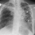
■ CXR
Should be performed if respiratory symptoms are present. Bronchial carcinoma may manifest as a hilar or peripheral opacity, cavitating mass, collapse of a segment of lung due to luminal obstruction, pleural effusion, elevated hemidiaphragm due to phrenic nerve palsy or destruction of an adjacent rib due to invasion. Lung abscesses present as a spherical shadow with a central lucency or air-fluid level. Bronchiectatic lungs have visibly dilated bronchi and multiple areas of consolidation. With fibrosing alveolitis, hazy shadowing of the lung bases produces a honeycomb appearance.
Specific Investigations
In view of the diverse nature of causes of clubbing, further investigations should be guided by the history and findings on clinical examination.
■ Urinalysis
Blood from microemboli in infective endocarditis.
■ Blood cultures (three sets in total: two sites at the same time and then one taken 12 hours later)
Infective endocarditis.
■ Echocardiography
Useful in the diagnosis of infective endocarditis, especially in the presence of large vegetations.
■ Sigmoidoscopy
Inflammatory bowel disease – characteristic findings include erythematous mucosa, areas of haemorrhage, contact bleeding and ulceration.
■ Rectal and colonic biopsy
Ulcerative colitis and Crohn’s disease.
■ Colonoscopy
Inflammatory bowel disease.
■ Barium enema
Inflammatory bowel disease.
■ Small bowel enema
Crohn’s disease.
■ Anti-tissue transglutaminase (ATA)
Coeliac disease.
■ Jejunal biopsy
Coeliac disease. May reveal subtotal villous atrophy reversible with a gluten-free diet.
■ CT thorax
Should be performed if bronchial carcinoma or thoracic empyema is suspected. Also useful to confirm the diagnosis of bronchiectasis and idiopathic pulmonary fibrosis.