Clinical practice

Published on 03/04/2015 by admin

Filed under Hematology, Oncology and Palliative Medicine

Last modified 22/04/2025

Print this page

rate 1 star rate 2 star rate 3 star rate 4 star rate 5 star
Your rating: none, Average: 0 (0 votes)

This article have been viewed 1177 times

42

Clinical practice

Red cell transfusion

Two questions need to be answered before transfusion of red cells is undertaken:

Some general indications for red cell transfusion are listed in Table 42.1.

Whole blood is now rarely used. Haemorrhage requires transfusion of fluids to maintain blood volume and red cells to raise the haemoglobin level. For correction of anaemia not responsive to other measures red cells stored in ‘optimal additive solution’ are used. There are few indications for red cells stored in plasma.

Practicalities of red cell transfusion

All those involved in the prescription and administration of blood should follow local guidelines with respect to patient identification and the checking of the compatibility and viability of the transfused units. Critical information is contained on the blood bag and the attached compatibility label (Fig 42.1). No discrepancies are permissible. Most serious adverse transfusion reactions are due to transfusion of the wrong blood to the patient (Fig 42.2). Errors can be reduced by newer technologies such as bar coding and radiofrequency chips – these generally rely on machine readable data on patient wristbands.
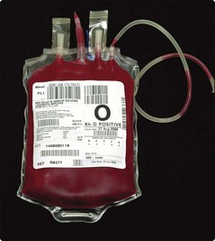
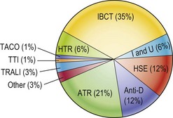
In shocked patients blood is transfused rapidly, the precise rate dependent on the monitoring of vital signs such as pulse, blood pressure and urine output. Transfusion for correction of anaemia is usually a more elective process. Units of red cells are typically given over 2–4 hours and a rise of around 10 g/L of haemoglobin can be expected from each unit. Red cells are infused via specially designed sterile ‘giving sets’ which contain 170 µm filters. Careful monitoring is particularly important during the first 10 minutes of each unit.

Complications of red cell transfusion

Immediate

Haemolytic transfusion reactions. These potentially fatal reactions arise from the transfusion of incompatible blood (usually for ABO). Symptoms often occur within minutes and may include chest, abdominal and loin pain, vomiting, a ‘burning’ skin, dyspnoea and headache. Common signs are fever, tachycardia and hypotension. Renal failure and disseminated intravascular coagulation (DIC) can follow. Once a haemolytic reaction is suspected, the transfusion should be stopped and the venous access used to give crystalloid. The transfused unit should be checked (is another patient about to get a ‘wrong’ unit due to a mix up?) and the blood bank informed. Initial investigations must include blood samples from the patient for a blood count and film, blood group, antibody screen and direct antiglobulin test. The blood bank will also repeat tests on the donated unit. Management of complications will require senior advice and often intensive care. The overall mortality of ABO incompatible transfusion is approximately 10%.

Non-haemolytic transfusion reactions. The majority of adverse reactions to blood are ‘febrile reactions’ caused by antileucocyte antibodies in the patient. Uncomplicated febrile reactions are simply managed by slowing the transfusion and giving paracetamol. Routine leucodepletion of red cells reduces such reactions. Occasionally patients develop allergic reactions with urticaria, wheezing and (rarely) anaphylaxis.

Transfusion associated circulatory overload (TACO). Care must be taken not to transfuse too rapidly, especially in elderly patients with heart disease.

Transfusion-related acute lung injury (TRALI). This is an acute syndrome occurring within 6 hours of transfusion and characterised by respiratory distress, hypoxia, bilateral pulmonary infiltrates and a fever. Donor antibodies to HLA class I and II antigens and/or granulocyte-specific antigens have been implicated in pathogenesis. Mortality is around 10%.

Delayed

Infection. Bacteria, viruses and parasites may all be transmitted via blood transfusion. Blood is screened for the relevant agents and in practice the greatest risk is of bacterial contamination. To help reduce the chance of transmission of the abnormal prion associated with variant Creutzfeldt–Jakob disease (vCJD) red cell donations are leucodepleted and plasma is increasingly imported from countries with no bovine spongiform encephalopathy (BSE). The significance of transmission of infection from blood can depend on the status of the recipient. Thus, cytomegalovirus (CMV) is of little relevance in healthy adults but potentially life-threatening in a patient receiving an allogeneic stem cell transplant or in a low birthweight premature infant.

Delayed transfusion reactions. These occur approximately 5–10 days after transfusion and are caused by a previously undetected antibody being boosted by transfusion of incompatible cells. Characteristic features include fever, jaundice and a falling haemoglobin. They are only rarely fatal.

Iron overload. A unit of blood contains around 250 mg of iron. Iron is only lost from the body in small amounts and repeated transfusion can lead to accumulation and toxic effects identical to those seen in haemochromatosis. Where repeated transfusion is predictable in a younger person (e.g. in thalassaemia), chelation of iron limits overload and prolongs life.

Massive blood transfusion

Massive transfusion is defined as replacement of the patient’s whole blood volume by stored allogeneic blood in less than 24 hours. There have been recent changes in practice driven by the military experience of trauma with increased early use of plasma and platelets. Problems can still arise in part due to the inevitable deficiencies of stored blood. Shortage of clotting factors and platelets in transfused blood may exacerbate haemorrhage. It is important to monitor haemostasis by checking the basic coagulation screen and replacing components accordingly. Metabolic disturbances are less common but include hyperkalaemia, hypocalcaemia, acidosis and citrate toxicity. Rapid transfusion can cause hypothermia; this can be minimised by carefully controlled blood warming.

Alternatives to allogeneic blood transfusion

Use of the patient’s own blood for transfusion rather than allogeneic blood minimises the risk of infection. Selected patients awaiting elective surgery can ‘pre-deposit’ blood in the weeks prior to the operation. An alternative approach, now more favoured, is to use specially designed equipment to ‘salvage’ blood lost during surgery and reinfuse it back into the patient. Other strategies to reduce blood transfusion include the active treatment of anaemia, strict application of transfusion triggers, anaesthetic measures or drugs (e.g. tranexamic acid) to reduce blood loss and biological agents such as erythropoietin. Questions remain regarding optimal transfusion practice and there is a need for more randomized controlled trials. Possible future developments include manufactured haemoglobin solutions and platelet substitutes.

Transfusion of platelets and granulocytes

Platelet transfusion

This is used to treat or prevent haemorrhage in patients with significant thrombocytopenia. It is more useful where platelets are low due to underproduction (i.e. marrow failure) or dilution than where thrombocytopenia is due to immune destruction as in immune thrombocytopenia (ITP). Platelets are collected either from routine blood donations or from a single donor by plasmapheresis. They should ideally be matched with the patient for ABO and Rhesus. The standard dose for an adult is either a single plasmapheresis donation or 4–6 pooled standard donations. Where repeated platelet transfusions are given, patients can become sensitised against class I HLA antigens absorbed onto the platelet surface with the result that they derive a lower increment in platelet count than would be predicted (‘platelet refractoriness’). In these cases, platelet donors matched with the recipient’s HLA class I type can be selected. Platelet transfusion can cause non-haemolytic reactions and can transmit infection as for red cells.

Transfusion of plasma and plasma products

A wide range of plasma products is available for therapeutic use:

image Fresh frozen plasma (FFP). Plasma is collected from whole blood or derived from plasmapheresis prior to rapid freezing. FFP contains the full range of coagulation factors and indications for use are shown in Table 42.2. The normal dose in an adult is one litre. FFP can transmit infection and cause immunological reactions – it is not suitable for volume expansion alone.

image Cryoprecipitate. This is prepared from FFP by slow thawing and separation of the resultant precipitate. It is rich in fibrinogen and may be useful in the treatment of DIC and management of massive blood transfusion.

image Factor VIII and IX concentrate. See pages 72–73.

image Albumin. This is produced by fractionation of pooled plasma. Solutions for clinical use include human albumin 4.5/5%, human albumin 20% and plasma protein fraction (PPF). Albumin solutions are used for the treatment of severe hypoproteinaemia, particularly when associated with a low plasma volume. Concentrated solutions can help produce a diuresis in hypo-albuminaemia (e.g. in hepatic cirrhosis).

image Immunoglobulins. These can be ‘specific’ and used in passive prophylaxis against a range of infections (e.g. varicella zoster, tetanus) or to prevent haemolytic disease of the newborn (anti-Rhesus D). ‘Non-specific’ immunoglobulins are used for passive prophylaxis against hepatitis A, treatment of hypogammaglobulinaemia and in selected autoimmune disorders (e.g. ITP).