Cyst, Clear

Published on 10/03/2015 by admin

Filed under Obstetrics & Gynecology

Last modified 22/04/2025

Print this page

rate 1 star rate 2 star rate 3 star rate 4 star rate 5 star
Your rating: none, Average: 0 (0 votes)

This article have been viewed 1593 times

Cyst, Clear

Synonyms/Description

Simple cyst
Clear ovarian cyst

Etiology

A simple ovarian cyst is a clear, thin-walled, unilocular cyst.

Premenopausal

Simple cysts up to 3 cm in maximal diameter are considered normal physiologic follicles in cycling women, and thus should not be reported as a cyst, particularly when the patient is midcycle. If a clear simple cyst does not resolve, it may be extraovarian, specifically paraovarian or paratubal. A persistent thin-walled and unilocular cyst may also represent a serous cystadenoma.

Postmenopausal

Small simple cysts up to 1 cm may be seen in up to 21% of postmenopausal women, and these are not considered clinically significant, even though the patient is no longer cycling. These less than 1 cm cysts typically should not be reported on an ultrasound examination and do not require follow-up.
In a study by Greenlee and colleagues, 2217 (14.1%) out of 15,735 women screened sonographically had one or more simple cysts on ultrasound examination. Among women without a cyst on the first screen, 8.3% had a new simple cyst 1 year later. There was no statistical difference in rate of subsequent ovarian cancer when those with simple cysts were compared with those without a cyst.

Ultrasound Findings

A simple and clear ovarian cyst is a thin, smooth-walled structure containing fluid with no internal echoes or solid areas. The cyst is usually round but can be oval, especially as it regresses and changes shape.
Any cyst that has a thick wall or any solid component is in a different category and discussed in other sections (refer to the differential diagnosis list for a complex cyst).
It is very important to scrutinize the sonographic appearance of cysts for any solid components or areas of wall thickening. A study by Valentin reports that 11 (0.96%) out of 1148 masses classified as unilocular cysts on ultrasound were malignant. However, postoperatively, 7 of the 11 malignancies thought to be unilocular cysts on ultrasound had gross papillary projections on the surgical specimen. Therefore accurate classification of a cyst as clear and unilocular is crucial on ultrasound examinations. Color Doppler may be useful to interrogate the walls of a cyst to better demonstrate any wall irregularity.

Differential Diagnosis

In a premenopausal patient, a simple cyst is most likely follicular and thus functional. If the patient is postmenopausal or the cyst does not regress in 3 months, the differential diagnosis includes cystadenoma, paratubal or paraovarian cyst, hydrosalpinx, or peritoneal inclusion cyst. Occasionally, serous cystadenomas can be unilocular and thin walled, although some may have a worrisome component such as a focally thick wall or septations. Paratubal or paraovarian cysts may mimic a follicle but can be distinguished sonographically by their location off the edge, or distant, from the ovary. Hydrosalpinges are rarely clear and thin walled, and are usually tubular with incomplete septations. Peritoneal inclusion cysts develop as a result of fluid being trapped within peritoneal adhesions. They typically have thin walls, an odd shape and may have multiple thin septations—all clues to the correct diagnosis.

Clinical Aspects and Recommendations

In general, simple clear cysts do not convey an increased risk of ovarian cancer, even in postmenopausal patients. With benign epithelial ovarian lesions there is no indication that malignant transformation occurs; for example, cystadenomas do not become cystadenocarcinomas. Current recommendations for management of simple cysts are the result of a collaborative consensus panel hosted by the Society of Radiologists in Ultrasound. The following recommendations pertain only to thin smooth-walled, completely clear, unilocular cysts.

In Premenopausal Women

Cysts less than 3 cm are considered normal and need not be followed. Cysts measuring 3 to 5 cm should be reported with a statement that they are almost certainly benign and may not need follow-up. Cysts between 5 and 7 cm are almost certainly benign but should be followed yearly with ultrasound. Cysts larger than 7 cm may be difficult to evaluate completely sonographically and might warrant further imaging.

In Postmenopausal Women

Cysts less than 1 cm are clinically inconsequential and may not be reported.
Cysts between 1 and 7 cm should be reported with a statement that they are almost certainly benign sonographically and yearly follow-up is recommended.
Cysts larger than 7 cm may be difficult to assess fully with ultrasound; thus surgical evaluation or alternative imaging should be considered.

Figures

image

Figure C4-1 Two views of a thin-walled unilocular cyst. This is a dominant follicle and is entirely normal. Such a cyst should not be mentioned in the ultrasound report because it is a normal finding.

image

Figure C4-2 Five-centimeter unilocular simple cyst showing the characteristic thin smooth walls and anechoic internal structure.

image

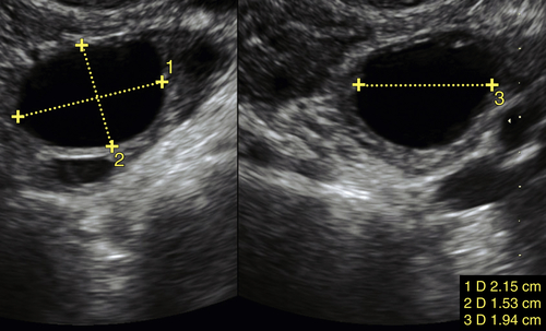
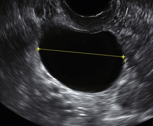
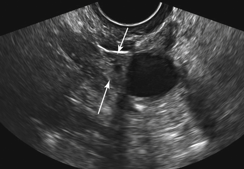
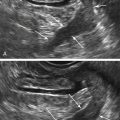
Figure C4-3 Differential diagnosis—paraovarian cyst (see Paratubal or Paraovarian Cysts). Note that the unilocular cyst is located adjacent to the ovary (arrows).


image

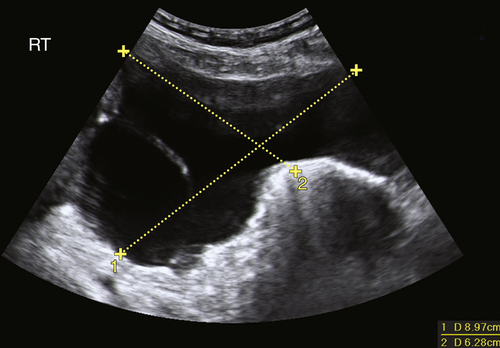
Figure C4-4 Differential diagnosis of large peritoneal inclusion cyst (see the section on complex cysts). This cyst is neither totally anechoic nor unilocular. It seems to take on the shape of the peritoneal space, a characteristic that is typical of a peritoneal inclusion cyst secondary to adhesions after pelvic surgery.


Suggested Reading

Greenlee R.T., Kessel B., Williams C.R., Riley T.L., Ragard L.R., Hartge P., Buys S.S., Partridge E.E., Reding D.J. Prevalence, incidence, and natural history of simple ovarian cysts among women >55 years old in a large cancer screening trial. Am J Obstet Gynecol. 2010;202  373.e1-9.

Levine D., Brown D.L., Andreotti R.F., Benacerraf B., Benson C.B., Brewster W.R., Coleman B., DePriest P., Doubilet P.M., Goldstein S.R., Hamper U.M., Hecht J.L., Horrow M., Hur H.C., Marnach M., Patel M.D., Platt L.D., Puscheck E., Smith-Bindman R. Society of Radiologists in Ultrasound. Management of asymptomatic ovarian and other adnexal cysts imaged at US Society of Radiologists in Ultrasound consensus conference statement. Ultrasound Q. 2010;26:121–131.

Valentin L., Ameye L., Franchi D., Guerriero S., Jurkovic D., Savelli L., Fischerova D., Lissoni A., Van Holsbeke C., Fruscio R., Van Huffel S., Testa A., Timmerman D. Risk of malignancy in unilocular cysts: a study of 1148 adnexal masses classified as unilocular cysts at transvaginal ultrasound and review of the literature. Ultrasound Obstet Gynecol. 2013;41:80–89.