[level-membership-for-cardiothoracic-surgery-category]Chapter 5
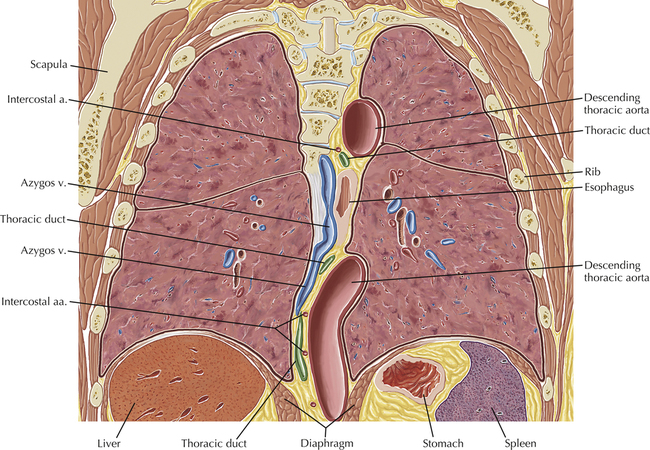
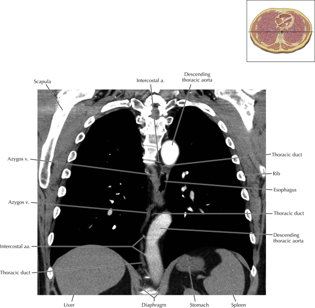
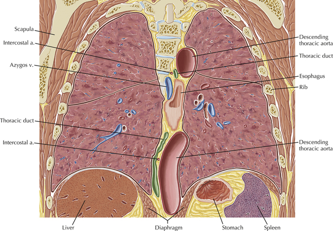
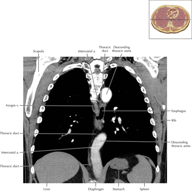
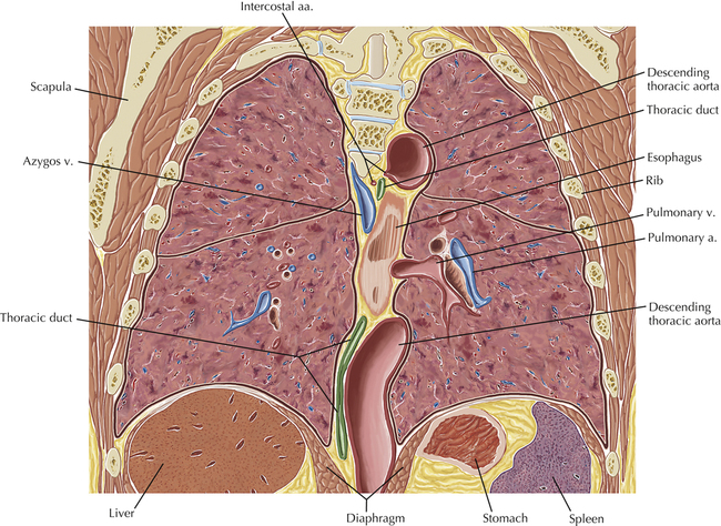
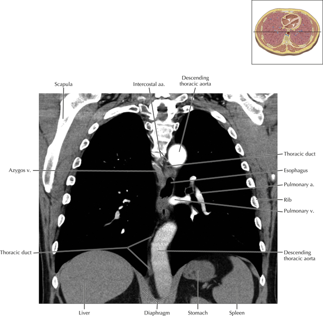
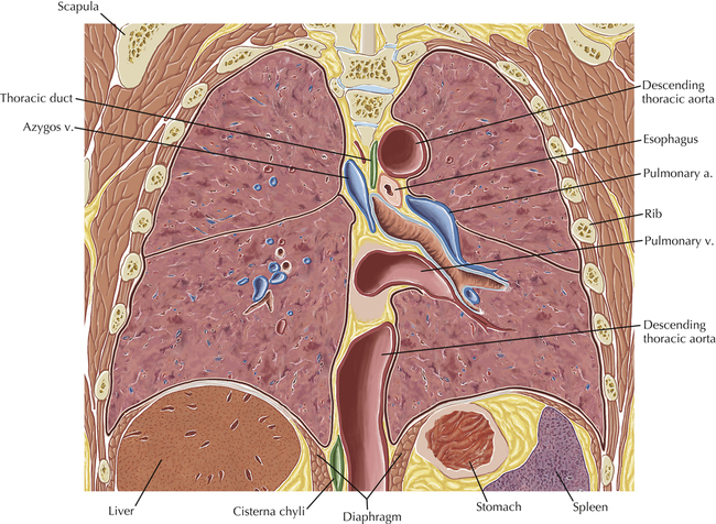
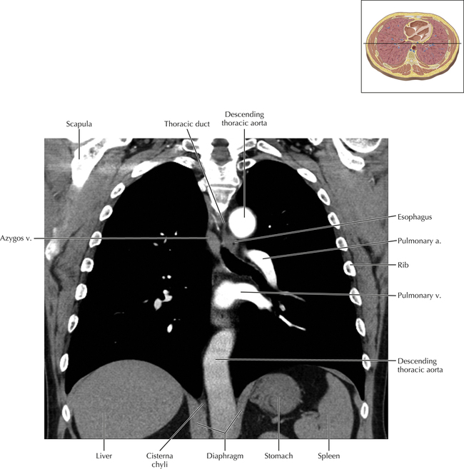
Cisterna Chyli and Thoracic Duct
[/level-membership-for-cardiothoracic-surgery-category][not-level-membership-for-cardiothoracic-surgery-category]Chapter 5
Cisterna Chyli and Thoracic Duct
Buy Membership for Cardiothoracic Surgery Category to continue reading. Learn more here
[/not-level-membership-for-cardiothoracic-surgery-category]
