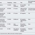
Circuit H
STATION 1
This station assesses your ability to elicit clinical signs:
CLINICAL SCENARIO
What further examination features would you wish to elicit at this point?
What additional information would you request at the end of the examination?
What investigation would you primarily request and what lesion do you expect to find?
STATION 3
This station assesses your ability to elicit clinical signs:
STATION 4
This station assesses your ability to elicit clinical signs:
INTRODUCTION
The examiner asks you to examine a 5-year-old child who presented to the ward with a cough.
CLINICAL SCENARIO
What clinical sign may be present on deeper examination of the mouth?
How will you present this information to the examiner?
What further aspects of the clinical examination are required?
What additional bedside tests would you perform?
Can you demonstrate to the examiner how to check for clubbing?
STATION 5
This station assesses your ability to elicit clinical signs:
STATION 6
This station assesses your ability to assess specifically requested areas in a child with a developmental problem:
INTRODUCTION
• Gross motor: You see spontaneous movements of all four limbs but with apparent spasticity bilaterally. She has poor head control. You ask her mother regarding her ability to roll, sit or stand and find that she will do none of them.
• Fine motor and vision: She does not reach for objects. She will hold toys placed in her hand but does not transfer them. There is no demonstrable pincer grip, though she has lost the grasp reflex. You note that she has bilateral coloboma of the iris. You test her ability to fix on your face and follow it to 90 or 180°. You do the same with a red toy and in both situations note that she is unable to follow. She appears to have a wandering gaze.
• Speech, language and hearing: You ask if the child has any language – noises, coos, babbles, words. The mother explains that she simply makes screams or non-specific noise. You ask if the child has had a hearing test and whether she seems to respond to noises by startling or quietening to her mother’s voice.
• Social, emotional and behaviour: You are unable to elicit any smiles or laughter and ask the mother if there have been any such actions noted by her. You note that the child is still in a nappy. The mother tells you she is fed orally but is fully dependent for toileting, though will cry when ‘dirty’.
What is the developmental age of this child in each of the areas of development?
STATION 7
This station assesses your ability to communicate appropriate, factually correct information in an effective way within the emotional context of the clinical setting:
STATION 8
This station assesses your ability to communicate appropriate, factually correct information in an effective way within the emotional context of the clinical setting:
BACKGROUND INFORMATION
His subsequent examination and developmental assessment were normal.
He had the following investigations:
• RAST to peanut 200 IU (negative to other nuts and milk)
• Skin test to peanut ++++ (negative to other nuts and milk).
What management plan would you recommend and would you prescribe the adrenaline (epinephrine) pen device (EpiPen)?
What additional follow-up would you arrange?
How could you check that the mother has understood your instructions?
STATION 9
This station assesses your ability to take a focused history and explain to the parent your diagnosis or differential management plan.
COMMENTS ON STATION 1
DIAGNOSIS: VENTRICULAR SEPTAL DEFECT REQUIRING MEDICAL/SURGICAL INTERVENTION
It is important when performing the cardiovascular exam to be thinking of what your findings imply as you go. The fact that the child is pink suggests that there is an acyanotic cardiac lesion. However, the evidence of poor growth and distress indicates the lesion is compromising. This child has a ventricular septal defect and must be examined for signs of failure. In this case it would be important to examine the abdomen for a palpable liver edge (and if present decide if it is pulsatile/smooth and determine the liver span by percussion) and then check that the femoral pulses are present. As the child has a thrill the murmur must be at least grade 4.
ASD (ostium secundum), also look for RSR in V1
Hypoplastic left heart
Left ventricular hypertrophy
Partial AVSD (ostium primum) with RSR in V1
Cardiac pacing
Complete AVSD (think Down’s)
ASD
Cor pulmonale
Eisenmenger’s reaction
VSD or AVSD
Aortic coarctation or stenosis
Hypertension.
A chest radiograph may be helpful in the diagnosis of congenital heart disease, and may be performed along with an ECG in hospitals with limited availability of echocardiography. The X-ray film should be analysed for:
1. Heart size and cardiothymic silhouette.
2. Size and prominence of the specific cardiac chambers and great arteries.
3. Pulmonary vascular markings and pulmonary plethora/oligaemia.
4. Associated abnormalities in bones/lungs/abdominal viscera (e.g. situs inversus, midline liver in asplenia/polysplenia, rib notching in older child with coarctation of the aorta, absent thymus in Di George).
Classical appearances in congenital heart defects include:
1. The ‘boot-shaped heart’ with oligaemic lung fields seen in cyanotic tetralogy of Fallot or some children with tricuspid atresia.
2. The ‘egg-shaped heart’ with plethoric lung fields and narrow pedicle in the cyanotic infant with transposition of the great arteries.
3. The ‘snowman in a snowdrift’ with plethoric lung fields and dilated SVC seen in supracardiac type of total anomalous pulmonary venous drainage (TAPVD).
COMMENTS ON STATION 2
DIAGNOSIS: BILIARY ATRESIA
• A breast-fed child less that 3 weeks old with a ‘yellow’ jaundice, normal stool and no other clinical finding is likely to have ‘breast milk jaundice’.
• A 2-month-old with mild ‘green’ jaundice, hepatomegaly, and a right hypochondrial incision may be post-Kasai procedure for biliary atresia.
• A 4-month-old with ‘green’ jaundice plus hepatomegaly, a central abdominal scar, abnormal umbilicus and a central line may well have TPN-induced conjugated jaundice (following surgery for neonatal necrotising enterocolitis or gastroschisis repair).
At the end of the examination in this case it is important to ask for the following:
| History | Gestation at birth |
| Birth trauma/cephalohaematoma | |
| Method of feeding | |
| Parenteral nutrition | |
| Significant family history | |
| Maternal and infant blood group | |
| Colour of urine and stool | |
| Examination | Dehydration |
| Dysmorphism, e.g. Alagille’s | |
| Bruising | |
| Pruritus | |
| Hepatomegaly | |
| Other features of note | |
| Inspect stool (e.g. pale stools and dark urine) | |
| Initial investigation | Full blood count and reticulocytes |
| Blood film | |
| Group and Coombs’ | |
| Packed cell volume | |
| Total and conjugated bilirubin | |
| Liver function test | |
| Urine: microscopy and culture | |
| Urinary bilirubin | |
| Further tests | G6PD assay |
| Urine metabolic screen | |
| Thyroid function | |
| TORCH | |
| Hepatic serology | |
| Investigating conjugated | Clotting |
| hyperbilirubinaemia (function) | Blood sugar |
| Investigating conjugated | Liver ultrasound |
| hyperbilirubinaemia (diagnosis) | HIDA scan |
| Liver biopsy | |
| α1-Antitrypsin | |
| Detailed endocrine investigation | |
| Bilirubin transport/conjugation defects | |
| Detailed metabolic investigation |
One would usually investigate a jaundiced infant at 2 weeks (if term and formula fed), 3 weeks (if pre-term or breast-fed) or as a matter of urgency if there is a history of jaundice accompanied by pale/grey/acholic stools and dark urine or the infant is systemically unwell (e.g. possible sepsis).
COMMENTS ON STATION 3
DIAGNOSIS: FRIEDREICH’S ATAXIA
Posterior column degeneration in the spinal cord is a feature of Friedreich’s ataxia.
The major clinical manifestations of Friedreich’s ataxia are progressive neurological dysfunction, cardiomyopathy and diabetes mellitus. The disease presentation is variable but early loss of joint position sense and vibration sense is typical. There is preservation of pain and temperature sensation. A progressive ataxia of all four limbs and gait occurs as a result of cerebellar dysfunction, often by 15 years of age.
The following pattern may be seen on examination:
• Cerebellar ataxia, dysarthria and nystagmus
• Wheelchair use (mean age of onset 11–25 years)
• Deep tendon reflexes are absent
• Plantar response is extensor due to pyramidal tract disease
• Pain/temperature sensation preserved
• Distal muscle atrophy (hands and feet)
• Skeletal abnormalities (pes cavus, hammer toes and progressive kyphoscoliosis)
• Visual impairment (due to optic atrophy) and swallow dysfunction
COMMENTS ON STATION 4
DIAGNOSIS: BRONCHIECTASIS; CLEFT LIP AND PALATE; HEARING IMPAIRMENT
’This 5-year-old boy looks comfortable at rest. I note he has bilateral hearing aids and previous facial surgery consistent with a repair of a congenital cleft lip and palate. He is clubbed in his fingers, consistent with chronic respiratory disease, and on examination of his chest I note that he has a portacath in situ on the left side and a scar in a similar position on the right side. He has diffuse, coarse breath sounds but no evidence of focal consolidation or effusion. The liver is not enlarged or displaced. My primary differential diagnosis is of bronchiectasis with associated bilateral conductive hearing impairment and a repaired cleft palate and lip.’
The standard bedside tests for the respiratory system should include the following:
REMINDER
| Mechanism | Causes |
|---|---|
| Respiratory | Bronchiolitis obliterans Severe asthma Previous severe pneumonia Pertussis |
| Genetic | Cystic fibrosis α1-Antitrypsin deficiency Primary ciliary dyskinesia (autosomal recessive) |
| Mechanical | Previous inhaled foreign body H-type tracheo-oesophageal fistula |
| Immune deficiency | Hypogammaglobulinaemia IgA deficiency |
| Idiopathic | |
COMMENTS ON STATION 5
DIAGNOSIS: TUBEROUS SCLEROSIS
The finding of adenoma sebaceum (angiofibromas) as described would make you consider the diagnosis of tuberous sclerosis.
Major features of tuberous sclerosis are:
• Facial angiofibromas or forehead plaques
• Periungual fibromas (outgrowth from nail-beds; don’t appear until puberty)
• Shagreen patch (an irregular area of connective tissue over the lumbar region)
• Ash leaf patches (hypomelanotic macules, i.e. areas of whiteness)
• Lymphangioleiomyomatosis and/or renal angiomyolipoma
• Cardiac rhabdomyoma (single or multiple; decrease in size with age)
Minor features of tuberous sclerosis are:
• Multiple randomly distributed pits in dental enamel
To show your understanding of how the disease may present with infantile spasms it may be useful to ask the mother how the child was first diagnosed and whether he has suffered with any seizures (present in 65% of cases). It may also show an understanding of the management of the condition if you ask about any extra help he may receive at home or in the classroom in light of special educational needs.
COMMENTS ON STATION 6
TESTS FOR INFANTS UNDER 6 MONTHS OF AGE
The Newborn Hearing Screening Programme (NHSP) is introducing hearing tests for all newborn babies in the UK within the first weeks of life.
1. Oto-acoustic emissions test: This is the most commonly used newborn screening test. A computer-generated ‘click’ is played through a small speaker in an earpiece placed in the child’s ear. A microphone (also in the earpiece) then listens for a soft echo, detectable if a healthy cochlea is present. The test is analysed by the computer and gives a ‘pass/fail’ result.
2. Automated auditory brain stem response test: This is also a screening test. The child has headphones placed over the ears and three small sensors are attached to the head. The child should be sleeping for the test. The headphones play a series of tones to the child, and a computer analyses the cerebral electrical responses. This tests both cochlear and auditory nerve function. A ‘pass/fail’ result is reported by the computer.
3. Auditory brain stem response test: If the screening test is failed, the child will be referred for this more detailed test. With a similar set-up to the automated test, the audiologist plays a series of sounds of different amplitudes and frequencies through the headphones while the sensors again transfer information to the computer. A record of the brain stem responses at the varying frequencies is provided and gives detailed information about the child’s hearing.
TESTS FOR INFANTS 6 MONTHS TO 2½ YEARS OF AGE
1. The distraction test: This is often known as the health visitor distraction test or the infant distraction test. It may be performed as a screening test, although it is being phased out with the introduction of newborn hearing screening. It is usually performed when the child is 7–9 months old. It requires two trained staff: the first to sit behind the child and make standardised test noises (such as the Manchester rattle, warbler, Nuffield rattle or a trained voice), varying them in frequency, while the second observes the child’s response to the sound. The reliability of this test is improved by it being performed in the audiology clinic. The child must be able to sit unsupported and have good head control.
2. Visual reinforcement audiometry test:
TESTS FOR CHILDREN 2½ YEARS OF AGE AND ABOVE
1. Speech discrimination test: The examiner tests the child’s ability to hear words at different volume levels without visual information.
2. Pure tone audiometry test (air and bone conduction): An audiometer produces pure sounds at certain frequencies and loudness, or vibrations at certain levels, and the child must respond with a button press or with a movement as part of a game.
3. Auditory steady-state evoked response test: This is similar to the automated auditory brain stem response test and involves the analysis of cerebral electrical activity monitored in response to sounds played into the ear by headphones.
VISION
The key principle with development is to work through a checklist of milestones in your mind and the approximate age of acquisition. If for each area of development you can reach a ‘passed milestone’ and a ‘failed milestone’ you can then build up an assessment of the child’s developmental age and present it to the examiner clearly.
| Age | Test or developmental level |
|---|---|
| Newborn | Check for red reflex (retinoblastoma), cataracts, ptosis, enlarged eyes (glaucoma) |
| 6 weeks | Expected to fix and follow faces/light for 45° |
| 8-12 weeks | Social smile and effective interaction, fix and follow for 180° |
| 6-8 months | Eyes should be aligned, only intermittent brief squint acceptable (e.g. when sleepy); hand regard, hands to mouth, directed reaching and transferring objects |
| 9-11 months | Grip development, recognise family versus strangers from face |
| 1 year | Pick up individual ’hundreds and thousands’ (pincer grip) |
| 2-3 years | Preferential looking or picture card tests for visual acuity, cover test for squint |
| 3 years | Sheridan-Gardner test (letter-matching test) |
| 4 years plus | Snellen chart (standardised test of visual acuity) |
COMMENTS ON STATION 7
The following would be appropriate:
You could continue by saying that Staphylococcus aureus is found to colonise 30% of the general population. MRSA is one type of this bacteria and can also be found on the skin of healthy people in the community, as well as colonising hospital populations. You can link on to talking about how it is routinely tested for when patients transfer from one hospital to another, mentioning those patients then have decontamination treatment of the skin and nose.
COMMENTS ON STATION 8
• Stridor due to laryngeal and pharyngeal oedema (tongue, lips and uvula)
Generally the management plan would be dependent on the reaction the child had suffered, the feelings of the mother and the services available to your hospital. In this case the APLS management of anaphylaxis needs to be understood. You would grade the severity of any reaction and give adrenaline (epinephrine) (or EpiPen), chlorphenamine, salbutamol and hydrocortisone or methylprednisolone as appropriate.
• Severe: Symptoms of airway compromise, shock, neurological signs, or severe gastrointestinal upset
• Moderate: Symptoms include respiratory symptoms (breathlessness, wheeze, chest tightness), dizziness, sweating, nausea, vomiting, abdominal pain
• Mild: Manifestations are limited to the skin only (urticaria, erythema, angioedema)
The EpiPen (http://www.epipen.com) is to be administered when a child has signs or symptoms of anaphylaxis. The dose of adrenaline (epinephrine) is given by the auto-injector through the clothes if necessary. You should follow the ‘instructions for use’ on the patient information leaflet.
COMMENTS ON STATION 9
The structure of the next 13 minutes is essential in order to cover the necessary topic areas. Below is a proposed approach.
You should get some essential background information, as you would with any new presentation:
• Problem list, teams involved in the child’s care
• Past medical or surgical problems
• Medication, allergies, immunisations
• Social arrangement and allied medical professional involvement
In terms of questions relating to diet you will be aiming to find out:
• What is the feed regimen (type of feed, volume, bolus feeds, overnight continuous feed)?
• Any nutritional supplements (e.g. Maxijul®)? How is it given?
• Are they giving anything orally?
• Have they kept a food diary?
• Has the child benefited from the gastrostomy?
• How do the carers feel about using the gastrostomy?
• Has the child vomited, had abdominal symptoms or had bowel upset?
In a ‘failure to thrive’ or ‘static weight’ scenario, you may divide causes into:
• Insufficient energy intake (inappropriate strength or volume feed)
• Inability to absorb feed/components (malabsorption from GI illness, pancreatic insufficiency, feed intolerance or reduced transit time)
• Excessive energy expenditure (coexisting illness, heart failure, sports).
Are there any other co-morbidities making the child have an increased energy expenditure?
What is the weight pattern (plot on the growth chart)?
What factors do the carers feel are responsible for the weight problem?
What do the carers feel should be the next step?
The examiner may raise specific issues with you regarding Rett’s syndrome.
The main features of the syndrome include:
• Deceleration of head growth (may be the first sign of Rett’s syndrome)
• Stereotypic hand movements (midline hand-wringing, hand-to-mouth movements, licking or repetitive grasping)
• Seizures (variety of types, 50% intractable)
• Gross motor dysfunction and ataxia
• Abnormal breathing patterns (apnoea and hyperventilation).