Circuit G
STATION 1
This station assesses your ability to elicit clinical signs:
STATION 2
This station assesses your ability to elicit clinical signs:
INTRODUCTION
On entering the room you are asked to examine the abdominal system of a 10-year-old boy.
STATION 3
This station assesses your ability to elicit clinical signs:
CLINICAL SCENARIO
On general inspection she is not dysmorphic. You examine in the following order:
• Acuity: Testing each eye individually (Snellen chart) – no defect.
• Visual fields: Testing each eye individually – no defect.
• Eye movements: You note that there is reduced lateral movement of the left eye. All other movements of both eyes are normal. The girl complains of diplopia on left lateral gaze. There is no nystagmus.
• Squint: You find that there is no tropia or phoria present.
• Pupils: The pupils are equal and reactive to light and accommodation.
• Fundoscopy: You find bilateral mild papilloedema (blurred disc margins and venous congestion) with no haemorrhage or exudates.
What nerve(s) are involved to give this pattern of external ophthalmoplegia?
What is the most likely cause of the vomiting?
STATION 4
This station assesses your ability to elicit clinical signs:
STATION 5
This station assesses your ability to elicit clinical signs:
INTRODUCTION
On entering the room you are invited to comment on the appearance of Crystal, a 6-year-old girl.
STATION 6
This station assesses your ability to assess specifically requested areas in a child with a developmental problem:
STATION 7
This station assesses your ability to communicate appropriate, factually correct information in an effective way within the emotional context of the clinical setting:
BACKGROUND
Dean was a normal term delivery and has a normal developmental history. He has no other medical problems and takes no other medication. He lives with his mother (a heavy smoker) and sister, both of whom suffer with eczema. He has a significant persistent night-time cough and daytime wheeze with exercise.
STATION 8
This station assesses your ability to communicate appropriate, factually correct information in an effective way within the emotional context of the clinical setting:
STATION 9
This station assesses your ability to take a focused history and explain to the parent your diagnosis or differential management plan:
COMMENTS ON STATION 1
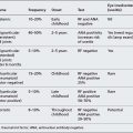
DIAGNOSIS: REPAIR OF AORTIC COARCTATION; TURNER’S SYNDROME
A thorough knowledge of cardiovascular defects, their management and their sequelae is vital for the exam. It is important that you know what the common scars look like – reading about a lateral thoracotomy scar is not the same as having seen one. In this case the small secondary scar is probably from a chest drain.
| Age | Clinical findings |
|---|---|
| Neonatal | Dorsal oedema of hands and feet |
| Redundant nuchal skin folds (secondary to in utero cystic hygromas) | |
| Low birth weight and reduced length | |
| 17-45% cardiac lesion (bicuspid aortic valve, coarctation of aorta, aortic stenosis, hypoplastic left heart) | |
| Developmental dysplasia of the hip (DDH) more common | |
| Childhood | Short stature (proportional) |
| 10% developmental delay | |
| Facial abnormalities (epicanthic folds, small mandible, prominent ears, high palate) | |
| Webbed neck | |
| Low posterior hairline | |
| Prominent ‘shield’ chest | |
| Widely spaced nipples | |
| Cubitum valgum | |
| Hyperconvex fingernails | |
| Grommits for ‘glue ear’ common | |
| Teenage | Failed onset of pubertal development (10% have breast enlargement) |
| Progressively more prominent pigmented naevi | |
| 30% renal abnormalities | |
| 70% impairment of non-verbal perceptual motor and visuospatial skills | |
| 15-30% hypothyroid | |
| Scoliosis, lordosis and kyphosis more common |
GENETIC ABNORMALITY
Turner’s syndrome occurs in 1 in 2500 to 1 in 3000 female live births. The most frequent genetic abnormality is monosomy X (45,X; 50%). The remainder of cases may have a duplication of the long arm of one X (46,X,i(Xq)) or a mosaicism (e.g., 45,X/46,XX). One to two percent of all conceptuses have 45,XO karyotype but over 99% will spontaneously abort.
COMMENTS ON STATION 2
DIAGNOSIS: LEUKAEMIA
In this station you have been presented with a Cushingoid 10-year-old with signs of anaemia, petechiae, alopecia and a central venous line.
The most likely reasons for a child having a central venous line are shown in the table below.
| Patient | Notes |
|---|---|
| Oncological | Needs chemotherapy |
| Haematological | Haemophilia requiring regular Factor VIII injections |
| Gastrointestinal | Total parenteral nutrition, e.g. short gut syndrome (secondary to Crohn’s disease or neonatal enterocolitis) |
| Renal | Red and blue ends for afferent and efferent lumens for haemodialysis |
| Immune deficient | Regular antibiotics or immunoglobulin replacement |
Presentation of these findings should be done succinctly; for example:
Note that the term ‘finger-breadths’ was used rather than an estimated span in centimetres; it will save you having to bring out a tape measure when the examiner challenges you! Paediatric oncology is an important subspecialty to which the candidate may not have been exposed; however, it is vital prior to the examination to have sought clinical experience in this area. In the communication skills station you could be required to ‘break the bad news’ to the parents of a child with newly diagnosed acute lymphoblastic leukaemia, while in the history-taking/management planning scenario you may have to discuss with patients and their families the common problems affecting their chronic care. You must be able to explain the side effects of long-term steroid use; the following mnemonic may help:
COMMENTS ON STATION 3
DIAGNOSIS: BENIGN INTRACRANIAL HYPERTENSION
| Category | Cause |
|---|---|
| Intracranial haemorrhage | Traumatic brain injury |
| • Extradural haematoma | Expanding arteriovenous malformation |
| • Subdural haematoma | Ruptured cerebral artery aneurysm |
| • Subarachnoid haemorrhage | |
| • Intracerebral haemorrhage | |
| Infections | Mastoiditis |
| Meningitis | |
| Encephalitis | |
| Vascular | Ischaemic infarcts |
| Vasculitis | |
| Neoplastic | Intracranial tumour (primary or metastatic) |
| Haematology | Sickling syndromes |
| Polycythaemia | |
| Prothrombotic states | |
| Other | Hydrocephalus |
| Benign intracranial hypertension | |
| Idiopathic |
What would be your first-line investigations?
• Bedside tests: blood pressure and urine dipstick analysis.
• Cranial imaging: preferably MRI or CT with contrast to exclude a mass lesion (essential prior to lumbar puncture).
• Formal ophthalmology opinion and visual field testing.
• Lumbar puncture with measurement of opening pressures.
• CSF sent for microscopy, culture, sensitivity, viral studies, glucose and protein.
• Full blood count, sickle cell screen, urea, electrolytes, calcium, coagulation screen and thrombophilia screen.
COMMENTS ON STATION 4
DIAGNOSIS: ASTHMA
| System | Conditions |
|---|---|
| Respiratory: extrathoracic These cause stridor (inspiratory noise) as the predominant symptom | Adenotonsillar hypertrophy Peritonsillar abscess Retropharyngeal abscess Epiglottitis Vocal cord dysfunction |
| Respiratory: intrathoracic | Tracheal stenosis or web Bacterial tracheitis Tracheo-bronchomalacia Tracheo-oesophageal fistula (H type or repair) Bronchiolitis (in the infant) Vascular ring |
| Respiratory: functional | Asthma Cystic fibrosis Recurrent aspiration Bronchopulmonary dysplasia (in the ex-pre-term infant) Bronchiectasis (including primary ciliary dyskinesia) |
| Cardiovascular | Cardiac failure (pulmonary oedema) Cardiomegaly |
| Gastrointestinal | Gastro-oesophageal reflux |
| Lymphoreticular | Mediastinal mass/lymphadenopathy Immunodeficiency |
What part of the respiratory system examination should you offer to examine next?
What additional bedside tests are important in this child?
In addition to the PEF you should ask to see the child’s sputum specimen pot (for the thick sticky green secretions seen in cystic fibrosis or bronchiectasis), measure a blood pressure and plot the child’s height and weight on the appropriate chart.
COMMENTS ON STATION S
DIAGNOSIS: NEUROFIBROMATOSIS TYPE 1 (NF1)
Diagnostic criteria for NF1 include the presence of at least two of the following:
2. Axillary or inguinal freckling
3. Lisch nodules (iris hamartomas) (> 2)
4. Neurofibromas (> 2) or one plexiform neurofibroma
5. Distinctive osseous lesion (kyphoscoliosis, sphenoid wing dysplasia, pseudoarthroses secondary to tibia/fibula bowing)
Differential diagnoses for conditions with café au lait patches include:
What systems will you now examine and how will you structure your examination?
• Skin: Examine all skin areas, including the axillae, groin and back.
• Cardiovascular: Full cardiovascular examination including blood pressure measurement, listening for renal artery bruits and palpating pulses.
• Neurological: Full neurological examination (including peripheral and cranial nerves, cognitive function and head circumference measurement).
• Eyes: Acuity, iris visualisation and fundoscopy.
• Musculoskeletal: Spine and limb examination, standing and sitting height.
COMMENTS ON STATION 6
DIAGNOSIS: GLOBAL DEVELOPMENTAL DELAY
The child in this case is actually 15 months old. She has developmental delay with truncal hypotonia. It is important not to make statements regarding the child’s true age if it is not known; instead refer to the developmental age.
What additional developmental reflexes could you describe or test in this child?
• Stepping reflex: With the child held in the vertical position their feet should be placed on the floor and then lifted lightly. This should encourage the child to make small stepping motions. This is slightly different from the placing reflex, where the feet are moved towards a small raised object. The feet should lift up to stand on that object. Both these reflexes are present at birth and disappear at approximately 6 weeks.
• Parachute reflex: The infant is moved rapidly face downwards towards a surface from a prone position. Both arms should spread out to break the fall. It appears at about 6 months but should definitely be present by 12 months. You support the child through the movement and don’t let go!
• Atonic neck reflex: The child must be supine and the head rotated to either side. The limbs on the side towards which the head is turned should both extend (with flexion of the contralateral limb). This reflex should disappear by 6 months.
COMMENTS ON STATION 7
This is bread-and-butter paediatrics and your examiner would expect you to perform to a high standard in order to pass the station. In order to do this you will need to have an understanding of asthma management in both acute and outpatient settings. Obviously, you must understand the advantages and disadvantages of interventions such as inhaled steroid. Also you must predict what the mother’s agenda is likely to be. What was the reason she didn’t want to start preventative treatment? This is a communication station so a regurgitation of facts about the British Thoracic Society (BTS) guidelines will be of no use if mum is concerned because her mother fractured her hip secondary to steroid use!
How would you initiate the conversation?
What do you expect the mother will want to know about the inhaled steroid?
| System | Side effects |
|---|---|
| Skin | Skin thinning, purpura, alopecia, striae, ‘Cushingoid facies’ |
| Eye | Cataracts, glaucoma |
| Cardiovascular | Hypertension, hyperlipidaemia |
| Gastrointestinal | Gastritis and ulceration, pancreatitis, bowel perforation |
| Renal | Fluid and electrolyte imbalance |
| Musculoskeletal | Myopathy, osteoporosis, avascular necrosis |
| Neurological | Hyperactivity, benign intracranial hypertension, euphoria |
| Endocrine | Diabetes mellitus, secondary adrenal insufficiency |
| Immune system | Increased opportunistic and standard infections |
In this case you will be able to reassure her that inhaled steroids at the standard dosing do not have the side effects seen in systemic corticosteroid therapy. Inhaled corticosteroids have been shown to cause oral candidiasis; there are case reports of adrenal suppression and short-term linear growth restriction but with attainment of normal adult height.
What would you include in an asthma plan for an acute exacerbation?
What would be the key issues for managing and monitoring background symptoms?
There is clear guidance from the BTS on the management of asthma. You should ensure that you have read and understood the latest edition of the guidelines (www.brit-thoracic.org.uk.)
The management of asthma should include the following:
• Minimise aggravating factors (e.g. smoking).
• Maximise patient concordance with treatment (e.g. by patient education).
• Age-appropriate treatment delivery (e.g. spacer device with mask for infants).
• Appropriate medication (as per ‘stepwise policy’).
• Appropriate referral to specialist care.
• Aim for minimal symptoms, minimise exacerbations, minimal intervention and normality of life.
COMMENTS ON STATION 8
This is a common scenario in life as a neonatal registrar, in interviews and exams. Remember that the station is targeted at assessing your communication skills and understanding of ethics rather than your in-depth understanding of neonatology.
What does resuscitation of a 24-week gestation newborn involve?
For this communication skills station it is useful to have worked on the neonatal unit and to have been involved in the resuscitation of extremely premature babies. For aspects of newborn life support (NLS) you should refer to the Resuscitation Council (www.resus.org.uk) course material. The following is a simple outline of the proceedings:
1. Experienced neonatal team in attendance at the delivery.
2. Neonatal ‘Resuscitaire’, equipment and drugs should be available.
3. Once the baby is born it is transferred to the Resuscitaire to be kept warm and to allow an initial assessment to be made (tone, colour, respiration, heart rate, response and, in the case of extreme prematurity, viability).
4. The baby is then intubated (prophylactic surfactant may be given), ventilation initiated and cardiac massage given with drug administration if appropriate.
5. The baby is transferred to the neonatal unit when stabilised.
In this scenario the following important points may be mentioned:
1. ‘It is sadly not possible for all babies born at this gestation to survive, and the initial resuscitation and first 24 hours are the most crucial.’
2. ‘The resuscitation of an extremely pre-term baby will involve a lot of people and the baby will require a tube being placed into its windpipe for us to give some medicine and to help him or her breathe.’
3. ‘If we are able to stabilise your baby we will take him/her to the neonatal unit where we will keep the baby warm, give fluids by drip and necessary medications.’
4. ‘Babies born this prematurely are poorly developed, in particularly the lungs and brain, and it is possible that the baby will have some long-term problems because of this fact.’
In the UK, morbidity and mortality data are available for neonates born prematurely through the Epicure study (bmj.bmjjournals.com/cgi/content/ full/319/7217/1093/DC1).
COMMENTS ON STATION 9
How will you structure the interview?
1. Initial introduction and clarification of reason for attending the clinic.
2. Period of active listening to ensure you have given the family a chance to tell you their concerns.
3. Directed questioning to ensure you have investigated your differential diagnoses appropriately with a detailed history of a typical ‘attack’.
What are your differential diagnoses for ‘loss of consciousness’ in this age group?
| System | Cause |
|---|---|
| Neurological | Seizure disorder Behavioural Reflex anoxic seizure Cerebrovascular accident |
| Respiratory | Respiratory arrest with hypoxia Cough syncope Breath-holding |
| Cardiovascular | Tachyarrhythmias (SVT, long Q-T) Bradyarrhythmias (AV conduction defects) Cardiomyopathy Vasovagal episodes Postural hypotension Shock, including anaphylaxis Left ventricular outflow obstruction, including aortic stenosis Congenital heart disease (including Fallot’s tetralogy) Pulmonary hypertension |
| Metabolic | Hypoglycaemia |
| Drug-induced | Inhaled nitrites Antihypertensive agents Tricyclic antidepressant Drugs of abuse (unlikely intentional at this age) |
What will your management plan involve?
For recurrent episodes that you feel are likely cardiac in origin you may suggest:
If you feel that the episodes are representing seizure activity, you could suggest: