Chronic Pancreatitis

Published on 19/07/2015 by admin

Filed under Radiology

Last modified 22/04/2025

Print this page

rate 1 star rate 2 star rate 3 star rate 4 star rate 5 star
Your rating: none, Average: 0 (0 votes)

This article have been viewed 4476 times

 Pancreatic parenchymal and intraductal calcification virtually diagnostic of chronic pancreatitis

image Pancreatic atrophy (often more apparent in body/tail)
image Fibroinflammatory “mass” related to chronic pancreatitis may be very difficult to differentiate from malignancy

– Most common in head and may demonstrate variable enhancement due to presence/absence of fibrosis
• MR: More sensitive for early changes compared to CT 

image Loss of normal high T1WI signal of parenchyma
image ↓ parenchymal enhancement on T1WI C+ arterial phase
image Dilated (> 3 mm), irregular pancreatic duct with strictures and dilated side branches (“chain of lakes” appearance)
image Stones within pancreatic duct appear as signal voids
image Secretin MRCP may identify earliest signs of CP

– Loss of normal duct distension (due to ↓ duct compliance) after secretin administration
– Secretin may improve visualization of side branches
– ↓ secretion of fluid into duodenum suggests pancreatic exocrine dysfunction

PATHOLOGY

• Most commonly caused by alcohol abuse (∼ 75% of cases)
• Other causes include idiopathic, hereditary pancreatitis, autoimmune pancreatitis, and systemic diseases
• Gallstones, hyperlipidemia, trauma, and drugs often cause acute/recurrent pancreatitis, but rarely chronic pancreatitis

CLINICAL ISSUES

• Endocrine and exocrine deficiencies due to progressive destruction of gland may lead to steatorrhea and diabetes
• Most patients treated with pain management, lifestyle modification (cessation of alcohol and smoking, frequent small meals), and pancreatic enzyme replacement
• Surgery an option in patients who fail medical therapy
image
(Left) Ultrasound image demonstrates multiple large, coarse calcifications image 
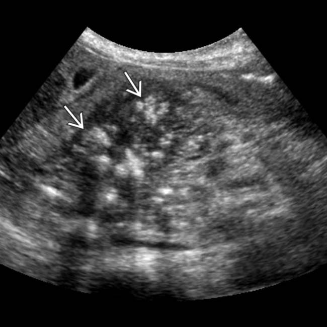
in the pancreatic head, some of which demonstrate posterior acoustic shadowing, compatible with chronic pancreatitis.

image
(Right) Coronal MRCP demonstrates a dilated main pancreatic duct with dilated side branch ducts image
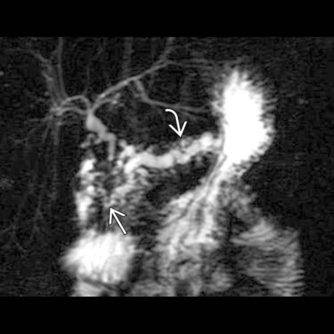
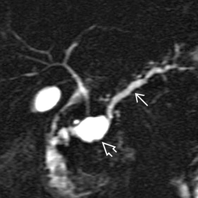
, as well as a distal pancreatic duct stricture image. Chronic pancreatitis is a scirrhous process that commonly causes stricture or occlusion of the ducts.
image
(Left) Coronal MRCP MIP reconstruction demonstrates characteristic changes of “big duct” chronic pancreatitis, including a dilated main pancreatic duct with dilatation of multiple side branches, stricture image 
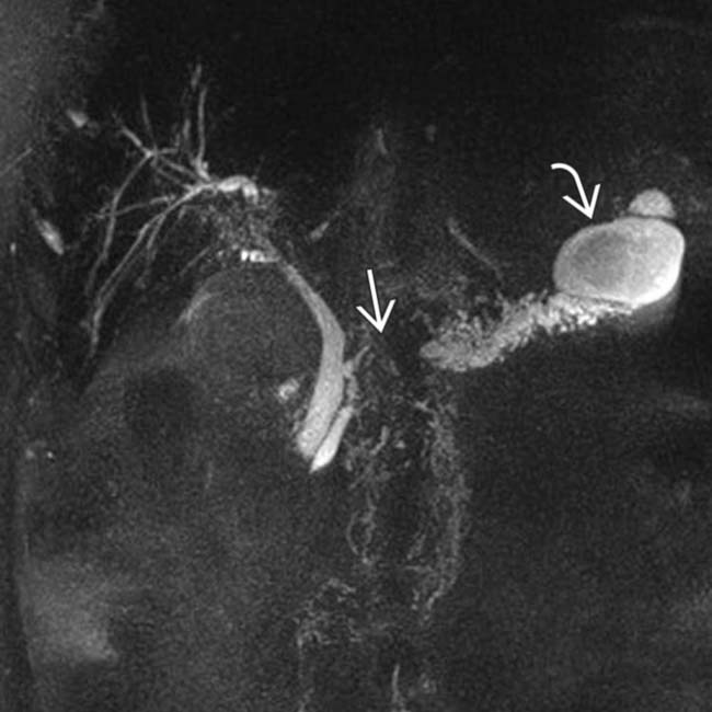
in the downstream duct, and a large pseudocyst image near the pancreatic tail.

image
(Right) Axial CECT demonstrates extensive parenchymal calcifications throughout the pancreas in a patient with known chronic pancreatitis. The multiple cysts image 
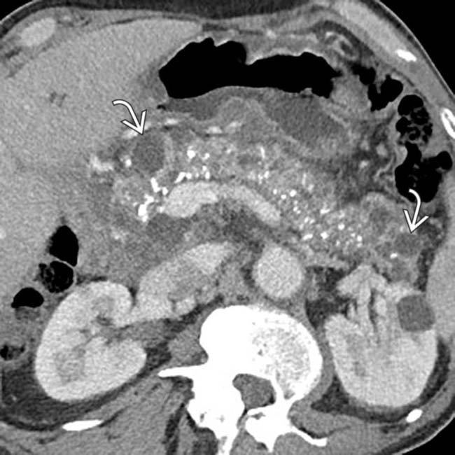
scattered throughout the pancreas in this case represent small pseudocysts.

TERMINOLOGY

Definitions

• Progressive, irreversible inflammatory damage to pancreas resulting in parenchymal fibrosis, morphologic changes, and loss of endocrine/exocrine function

IMAGING

General Features

• Best diagnostic clue

image Atrophic pancreatic parenchyma with a dilated, beaded main pancreatic duct (MPD) and intraductal calculi
• Size

image Pancreas usually atrophic
• Morphology

image Pancreatic calcification

– Almost diagnostic of chronic pancreatitis
– ∼ 90% of calcific pancreatitides are caused by alcoholism

image Other 10% = mostly hereditary pancreatitis
– Present in 40-60% of patients with alcoholic pancreatitis
• Other features

image 75% of cases in USA are due to alcoholism
image Developing countries: Malnutrition and alcoholism

CT Findings

• Earliest stages may not produce visible changes, but morphologic abnormalities more apparent in later stages

image Dilated, beaded, irregular pancreatic duct with strictures
image Pancreatic parenchymal and intraductal calcification virtually diagnostic of chronic pancreatitis (CP)

– ∼ 90% of calcific pancreatitis caused by alcoholism (remaining 10% mostly hereditary pancreatitis)
image Pancreatic atrophy (often more apparent in body/tail)
image Intra- and peripancreatic pseudocysts
• Splenic vein often thrombosed with resultant varices and splenomegaly
• Fibroinflammatory “mass” related to chronic pancreatitis may be very difficult to differentiate from malignancy

image Most common in pancreatic head and may demonstrate variable enhancement 

– Hypoenhancing mass usually due to fibrosis, whereas isodense mass implies lack of fibrosis

MR Findings

• Normal MR appearance of pancreas

image Parenchyma diffusely high signal on T1WI (≥ liver)
image Parenchyma variable in signal on T2WI
image Pancreas enhances avidly and homogeneously on T1W C+ images (hyperintense to liver on arterial phase and isointense on delayed phase)
image Normal pancreatic duct measures < 3 mm and side branches are not normally visualized
• More sensitive for early changes of chronic pancreatitis compared to CT (although less sensitive for calcifications)

image Loss of normal high T1WI signal of parenchyma (due to fibrosis replacing parenchymal proteinaceous fluid)
image Diminished parenchymal enhancement on T1W C+ images on arterial phase with increased delayed enhancement due to fibrosis
image ↓ parenchymal enhancement and T1WI signal can also be seen in older patients with age-related fibrosis
• Changes in pancreatic duct (usually later finding) nicely demonstrated on T2WI or MRCP

image Dilated (> 3 mm), irregular pancreatic duct with strictures and dilated side branches (“chain of lakes” appearance)
image Visualization of side branches, which are not normally visible, may be subtle sign
image Stones within pancreatic duct appear as signal voids
image Cambridge criteria for ERCP may be applied to MRCP
• Pancreatic atrophy and pseudocysts in later stages
• Secretin MRCP can help visualize earliest findings of chronic pancreatitis and evaluate pancreatic exocrine dysfunction

image Loss of normal duct distension (due to ↓ duct compliance) after secretin administration in CP

– Normal duct dilates ≥ 1 mm compared to baseline (< 1 mm distension suggests CP)
image Secretin may improve visualization of side branches (finding that suggests CP)
image ↓ secretion of fluid into duodenum suggests pancreatic exocrine dysfunction, suggesting CP

– Grade I: Fluid seen in duodenal bulb (most suggestive of pancreatic exocrine dysfunction)
– Grade II: Fluid seen in 2nd portion of duodenum
– Grade III: Fluid reaches 3rd portion of duodenum
• MR, like CT, cannot reliably distinguish fibroinflammatory mass (due to CP) from malignancy

Radiographic Findings

• Radiography

image Abdominal radiographs: May demonstrate small, irregular, or coarse calcifications (local or diffuse) in expected location of pancreas in upper abdomen
image Upper GI series: May reveal changes in 2nd part of duodenum

– Thickened, irregular mucosal folds, luminal narrowing, and varying degrees of atony with dilatation of proximal duodenum ± stomach
– Enlarged major duodenal papilla
• ERCP

image Considered gold standard test for chronic pancreatitis
image Dilated, irregular, and beaded main pancreatic duct with sites of stricture and dilated side branches
image Intraductal calculi appear as filling defects within MPD
image Intrapancreatic portion of CBD may be narrowed, but demonstrates smooth, tapered narrowing (not abrupt narrowing as with malignancy)

– May produce double duct sign due to stricture of distal CBD and MPD (similar to malignancy)
image Cambridge criteria for chronic pancreatitis on ERCP

– Normal: Normal MPD and side branches
– Equivocal: Normal MPD with < 3 abnormal side branches
– Mild: Normal MPD with ≥ 3 abnormal side branches
– Moderate: Abnormal MPD and ≥ 3 abnormal side branches
– Severe: Abnormal MPD with large cavity (> 10 mm), ductal obstruction, filling defects, or severe dilatation/irregularity and ≥ 3 abnormal side branches

Ultrasonographic Findings

• Grayscale ultrasound

image Not reliable means of diagnosis as pancreas often obscured by bowel gas
image Dilated MPD (± CBD) with echogenic parenchymal and ductal calcifications (with posterior acoustic shadowing)
image Gland may appear hypoechoic and heterogeneous
• Endoscopic ultrasound

image Multiple criteria for diagnosis including parenchymal abnormalities (hyperechoic foci, hyperechoic strands, lobulated parenchymal contour, cysts) and ductal abnormalities (dilated MPD, irregular MPD, hyperechoic duct walls, visible side branches, calcifications)

– Specificity ↑ as more criteria are present

Angiographic Findings

• May identify pseudoaneurysm of gastroduodenal or other arteries

Imaging Recommendations

• MR/MRCP (with secretin) is best noninvasive imaging test
• Endoscopic ultrasound may be helpful for early-stage disease and ERCP utilized primarily for intervention (stone extraction, stent, etc.)

DIFFERENTIAL DIAGNOSIS

Pancreatic Ductal Carcinoma

• Poorly marginated hypoenhancing mass with upstream parenchymal atrophy and abrupt obstruction of MPD/CBD
• Posterior extension into retroperitoneum with encasement and narrowing of mesenteric vasculature
• ERCP: Irregular, nodular, rat-tailed eccentric obstruction of pancreatic duct
• > 70% of patients present with advanced local disease and distant metastases
• Some cases of fibroinflammatory mass due to chronic pancreatitis and pancreatic cancer are impossible to differentiate without surgical resection and histology

Pancreatic Intraductal Papillary Mucinous Neoplasm (IPMN)

• Involvement of main pancreatic duct may simulate chronic pancreatitis clinically and on CT/MR
• Dilated MPD (± side branches) with parenchymal atrophy
• ERCP can more easily make distinction with ability to visualize mucus ± polypoid lesions within MPD

Senescent Change

• Pancreatic parenchyma atrophies with age (usually > 70 years), duct may mildly dilate, and parenchyma may demonstrate small foci of calcification
• MR may demonstrate ↓ parenchymal enhancement and T1WI signal in age-related fibrosis (like chronic pancreatitis)
• Degree of ductal dilatation usually quite mild without irregularity, strictures, or intraductal stones

PATHOLOGY

General Features

• Etiology

image Chronic pancreatitis most commonly caused by alcohol abuse (∼ 75% of cases in USA)

– Persistent, heavy alcohol consumption for > 10 years usually required to develop chronic pancreatitis
– Other causes include idiopathic (10-30% of cases), hereditary pancreatitis, tropical pancreatitis, autoimmune pancreatitis, and systemic diseases (most notably cystic fibrosis)
– Obstructive chronic pancreatitis due to narrowing/stricture of duct

image Includes congenital abnormalities that predispose to CP, including pancreas divisum, annular pancreas, and sphincter of Oddi dysfunction
– Toxins (tobacco) may play causative role
image Gallstones, hyperlipidemia, trauma, and drugs often cause acute/recurrent pancreatitis, but rarely CP
image Pathogenesis still debated but probably due to chronic reflux of pancreatic enzymes, bile, and duodenal contents with increased ductal pressure  

– Resultant activation of pancreatic stellate cells by toxins sets up fibroinflammatory response
• Genetics

image Hereditary pancreatitis may be related to several genes, including PRSS1 -cationic trypsinogen, SPINK1 -trypsin inhibitor, and CFTR

– Most commonly autosomal dominant trait with incomplete penetrance
– Can injure pancreas without other “toxins”
image Genetic predisposition toward pancreatitis related to alcohol: Some patients suffer irreversible injury with modest ingestion of alcohol
• Anatomic predispositions

image Pancreas divisum: Minor papilla opening may be inadequate to drain pancreatic secretions
image Annular pancreas: Pancreatic ductal obstruction and stasis of secretions

Gross Pathologic & Surgical Features

• Hard, scirrhous, atrophic pancreas with intraductal calculi and dilated MPD
• Multiple parenchymal calcifications
• Pseudocysts may be seen

Microscopic Features

• Atrophy and fibrosis of acini with dilated ducts
• Mononuclear inflammatory reaction
• Occasionally squamous metaplasia of ductal epithelium

CLINICAL ISSUES

Presentation

• Most common signs/symptoms

image Recurrent or chronic midepigastric pain (typically radiates to back and is worse after eating)
image Endocrine and exocrine deficiencies due to progressive destruction of gland and stricture of CBD: Steatorrhea, diabetes mellitus, and jaundice

– Usually clinically apparent only after 90% of gland function is destroyed
– Morphologic changes on imaging correlate with endocrine/exocrine dysfunction

image “Big” duct disease with gross abnormalities of MPD often results in steatorrhea and diabetes
image “Small” duct disease with normal MPD and relatively normal-appearing pancreas unlikely to produce steatorrhea and diabetes
• Clinical profile

image Patient with history of chronic alcoholism, recurrent attacks of midepigastric pain radiating to back, jaundice, steatorrhea, and diabetes
• Lab data

image Amylase and lipase not reliable for CP, as they may be normal or only minimally elevated
image Increased blood glucose levels and elevated fecal fat (diabetes and steatorrhea)
image Secretin test: Decreased amylase and bicarbonate

Demographics

• Age

image Usually middle-aged adults
image Hereditary pancreatitis may present < 20 years
• Gender

image M > F

Natural History & Prognosis

• Complications

image Diabetes and malabsorption
image Jaundice due to biliary obstruction
image Duodenal obstruction
image GI bleeding from varices (± gastritis) due to splenic vein or portal vein thrombosis
image Significant ↑ in pancreatic cancer incidence
• Prognosis

image Poor prognosis due to difficulty in relieving chronic pain and other symptoms

Treatment

• Most patients treated with pain management, lifestyle modification (cessation of alcohol and smoking, frequent small meals), and pancreatic enzyme replacement
• Surgery an option in patients who fail medical therapy

image Patients with “big” duct disease and a grossly dilated duct may undergo surgical decompression of duct (usually anastomosis of duct with Roux limb)
image Surgical resection (Whipple procedure, distal pancreatectomy, total pancreatectomy) is an option in patients with “small” duct disease

DIAGNOSTIC CHECKLIST

Consider

• Differentiate from other causes of MPD dilatation and glandular atrophy, including main duct IPMN and pancreatic adenocarcinoma
• Fibroinflammatory mass related to chronic pancreatitis may be very difficult to distinguish from pancreatic adenocarcinoma

Image Interpretation Pearls

• Glandular atrophy with dilated MPD, ductal calculi, thickened peripancreatic fascia ± pseudocyst are most telling signs for chronic pancreatitis
image
(Left) Axial CECT in a patient with chronic pancreatitis demonstrates an infiltrative hypodense pancreatic mass image
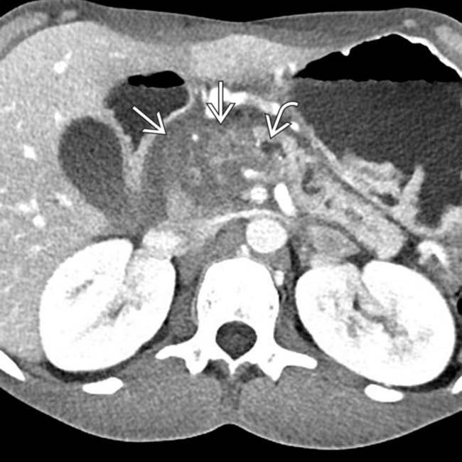
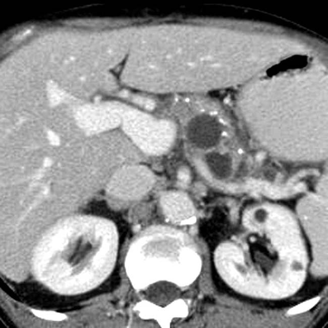
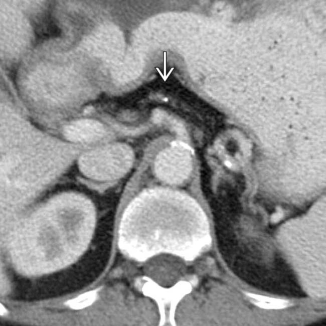
, with a dilated upstream pancreatic duct image. A few calcifications were present in the parenchyma (not shown). The patient underwent Whipple procedure due to concern for malignancy, where this was found to be a fibroinflammatory mass related to chronic pancreatitis.

image
(Right) ERCP shows irregular dilatation of the main pancreatic duct and side branches characteristic of chronic pancreatitis.
image
(Left) Coronal MRCP MIP reconstruction in a patient with chronic abdominal pain demonstrates subtle dilatation of a few scattered pancreatic duct side branches image
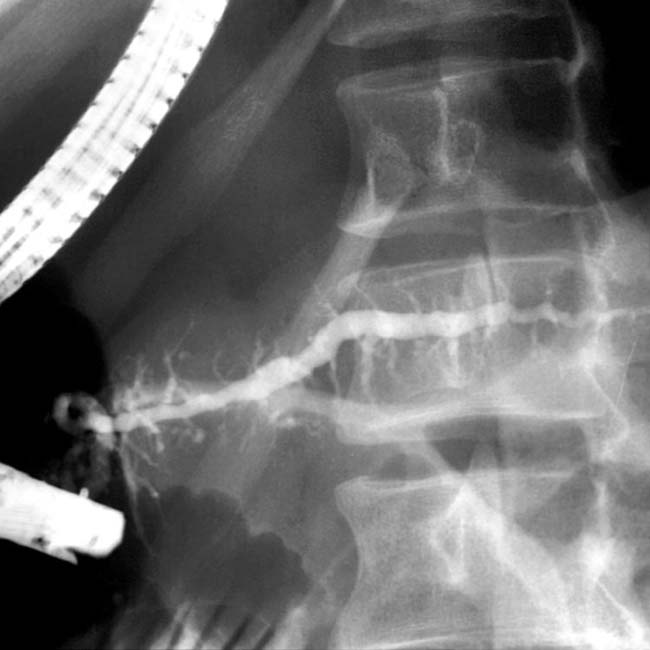
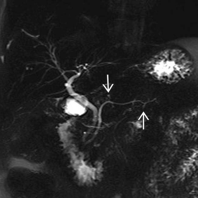
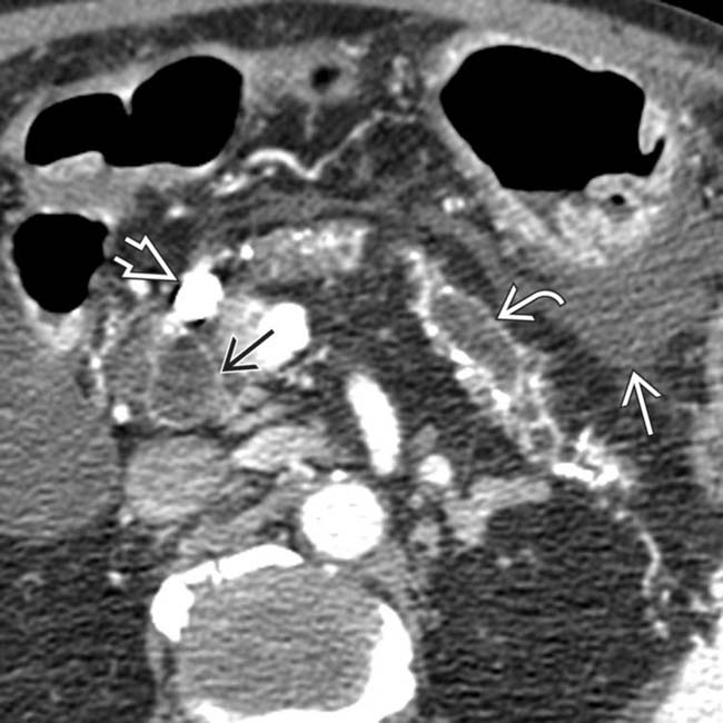
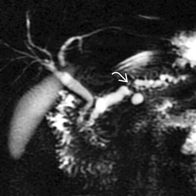
. Side branch dilatation can be an early sign of chronic pancreatitis, subsequently confirmed in this patient using endoscopic ultrasound.

image
(Right) Axial CECT shows a large, discrete stone image 
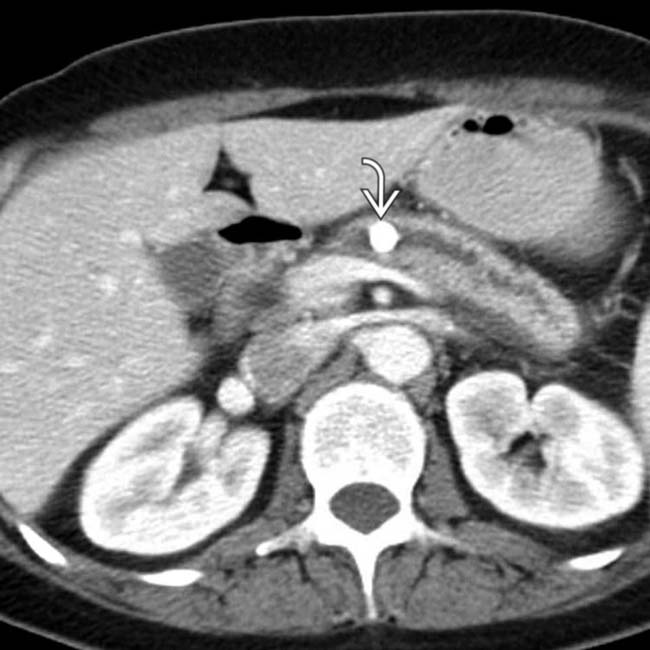
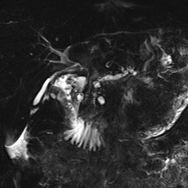
within a dilated main pancreatic duct in a patient with known alcohol-related chronic pancreatitis. The duct itself appears subtly irregular and beaded.
image
(Left) Coronal curved planar reformation from a CECT shows dilatation of the pancreatic duct image 
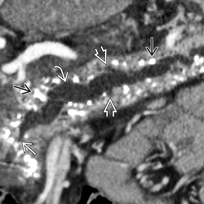
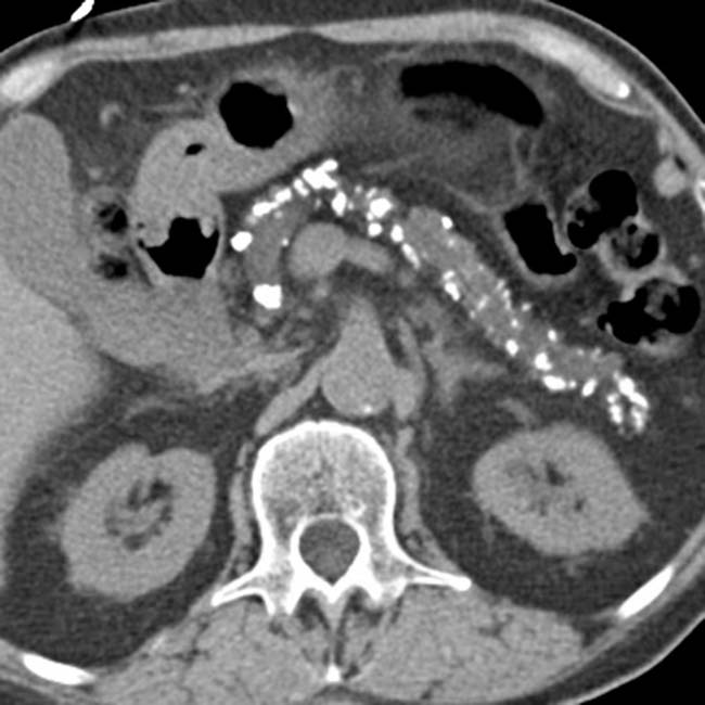
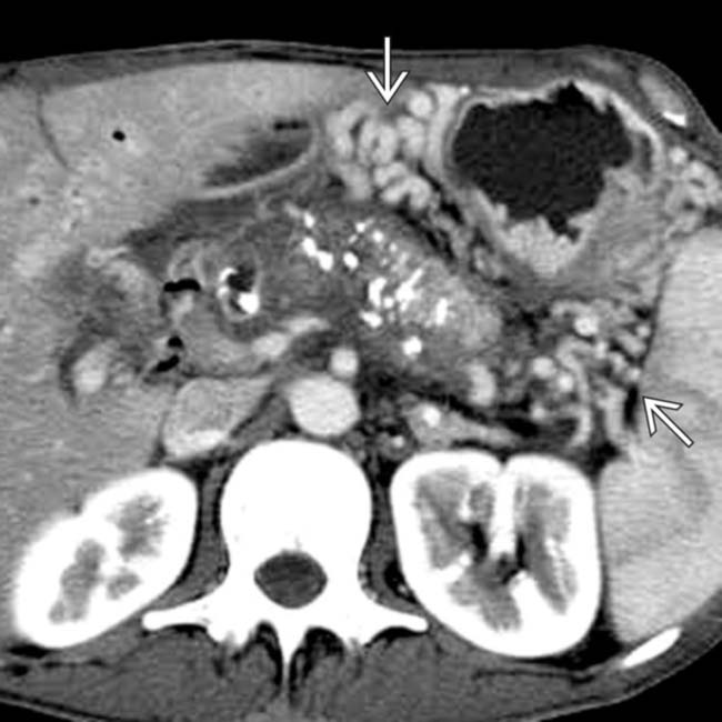
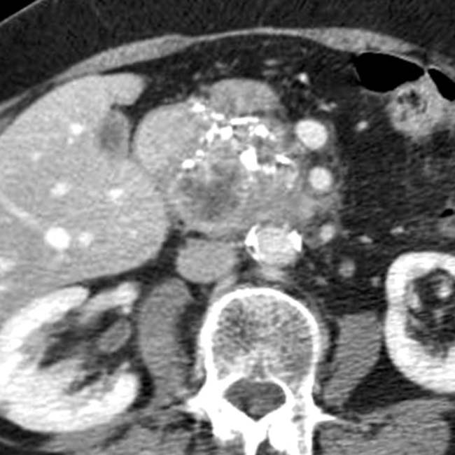
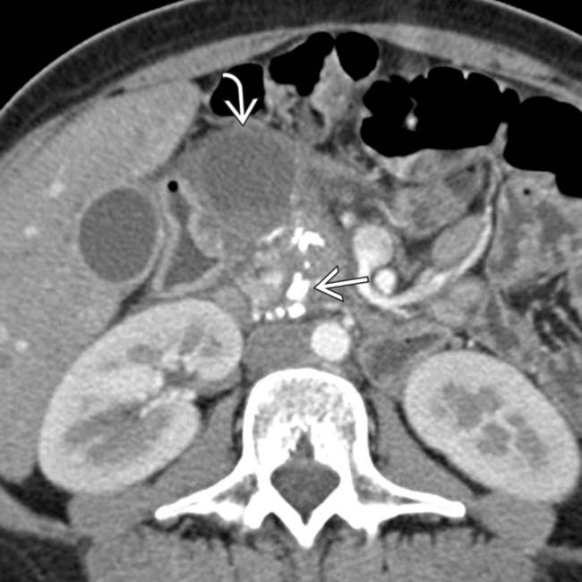
upstream from a stricture image in the head of the pancreas. Note the dilated side branches of the pancreatic duct image and extensive parenchymal calcifications image, diagnostic of chronic pancreatitis.

image
(Right) Axial MIP image from a CECT in the same patient illustrates extensive parenchymal calcifications image
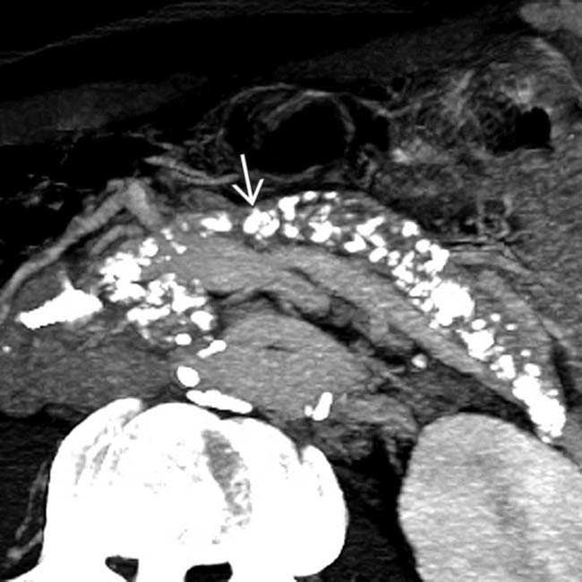
. This patient required enzyme replacement therapy due to exocrine insufficiency.
image
Axial CECT shows glandular atrophy, a dilated main pancreatic duct, and intraductal calculi.

image
Axial CECT shows parenchymal atrophy, calcifications, and small pseudocysts.
image
Axial CECT shows pancreatic calculi and perisplenic and perigastric varices image 
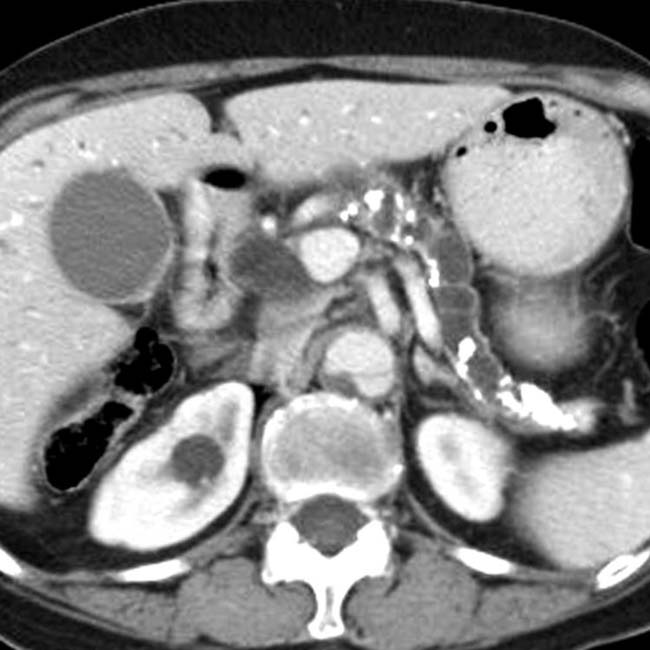
due to obstruction of the splenic vein.
image
MRCP shows long tapered narrowing of the common bile duct image 
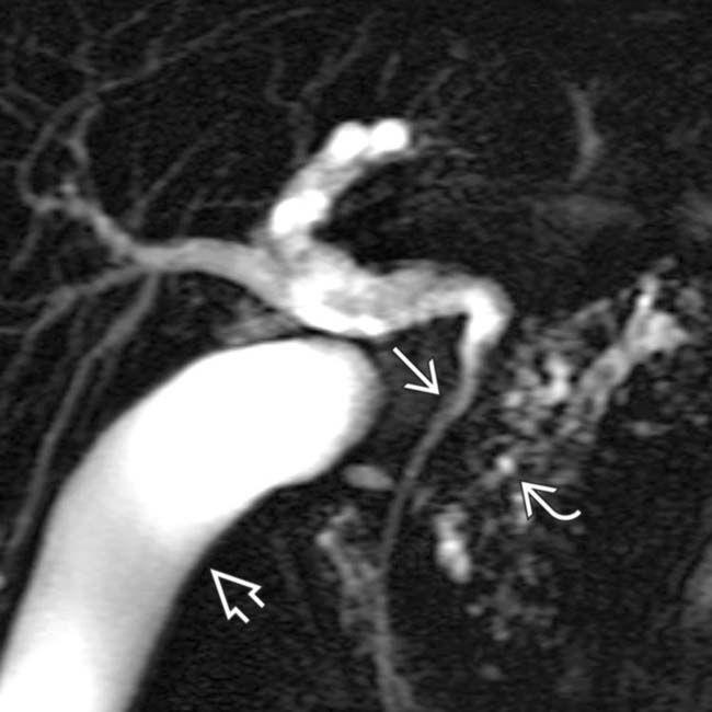
as it passes through the head of the pancreas. Note the irregular strictures and dilatation of the pancreatic duct image. The gallbladder is also seen image.
image
MRCP shows a dilated main pancreatic duct image and adjacent pseudocyst image.
image
Axial CECT shows marked parenchymal atrophy with fatty replacement and a dilated main pancreatic duct containing calculi image.
image
Axial CECT in a patient with chronic pancreatitis shows a large heterogeneous mass in the pancreatic head with parenchymal calculi. No tumor was found after surgical resection.
image
Upper GI series film from a patient with chronic pancreatitis shows marked narrowing of the lumen of the 2nd duodenum image with some fold thickening of the 3rd duodenum. The stomach was slow to empty.
image
Transverse grayscale ultrasound of the pancreas demonstrates a dilated main pancreatic duct image 
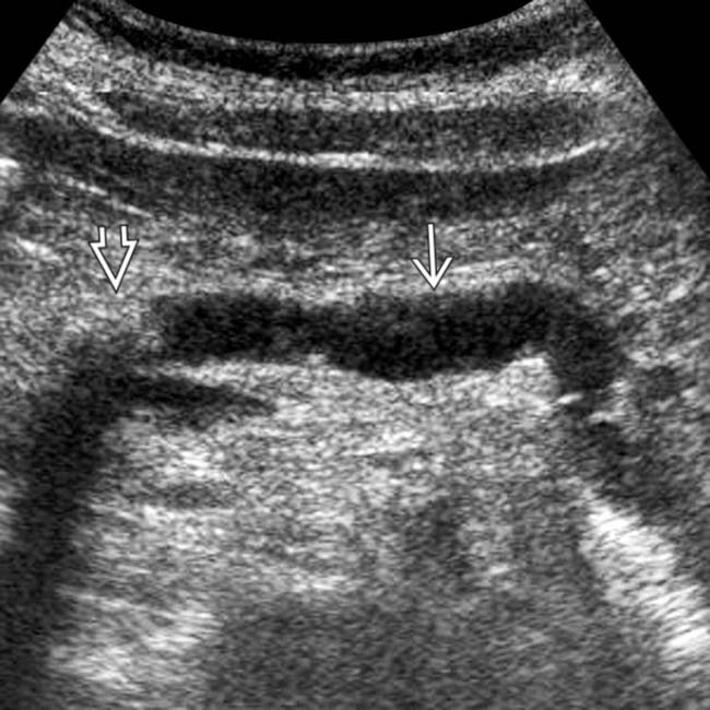
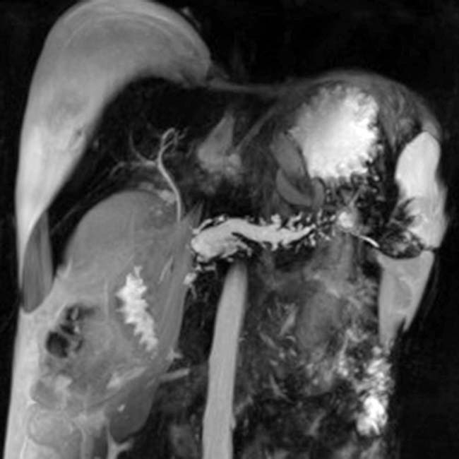
upstream from an obstructing intraductal stone image.
image
Axial CECT in the same patient shows marked parenchymal atrophy and dilatation of the main pancreatic duct image. Note the large intraductal stone image in the neck of the pancreas as well as a dilated common bile duct image. There is fluid in the lesser sac image from acute pancreatitis. This is an example of acute and chronic pancreatitis.
image
Axial CECT shows cavernous transformation image 
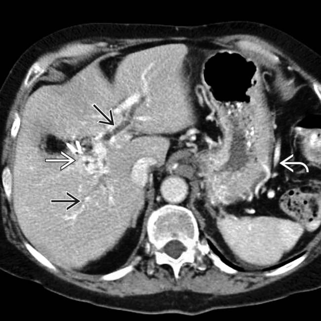
of an occluded portal vein with collateral veins in the porta hepatis and around the stomach image. The intrahepatic bile ducts are dilated image due to a stricture of the common bile duct as it traverses the pancreas.
image
Axial CECT in the same patient shows marked atrophy of the pancreas image
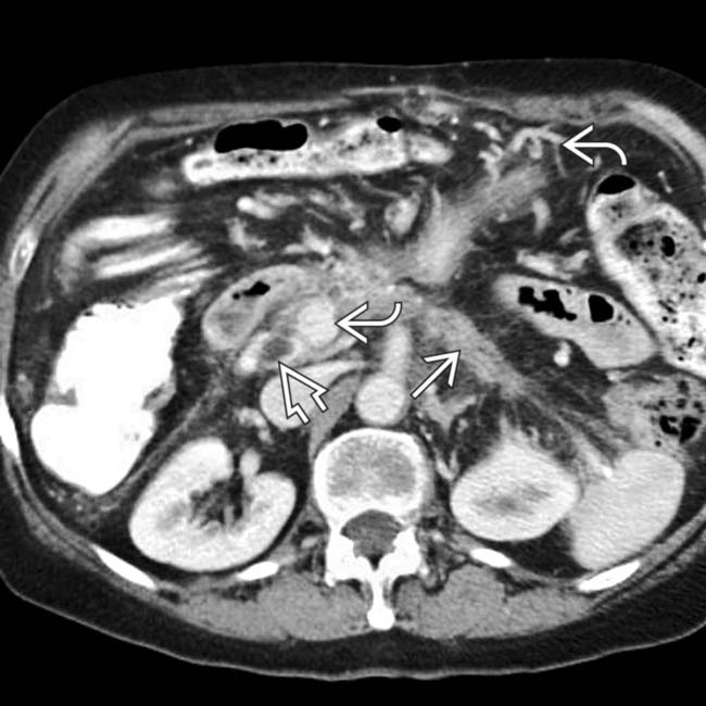
, dilation of the distal pancreatic duct image, and varices image within the pancreas and mesentery due to splenic and portal vein thrombosis.
image
Coronal MRCP of a patient with chronic pancreatitis causing bile duct stricture shows a long tapered narrowing of the intrapancreatic portion of the common bile duct image 
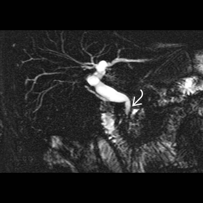
with dilation of the intrahepatic ducts.
image
ERCP of a 47-year-old woman with chronic pancreatitis due to alcohol abuse shows a cluster of calcifications image 
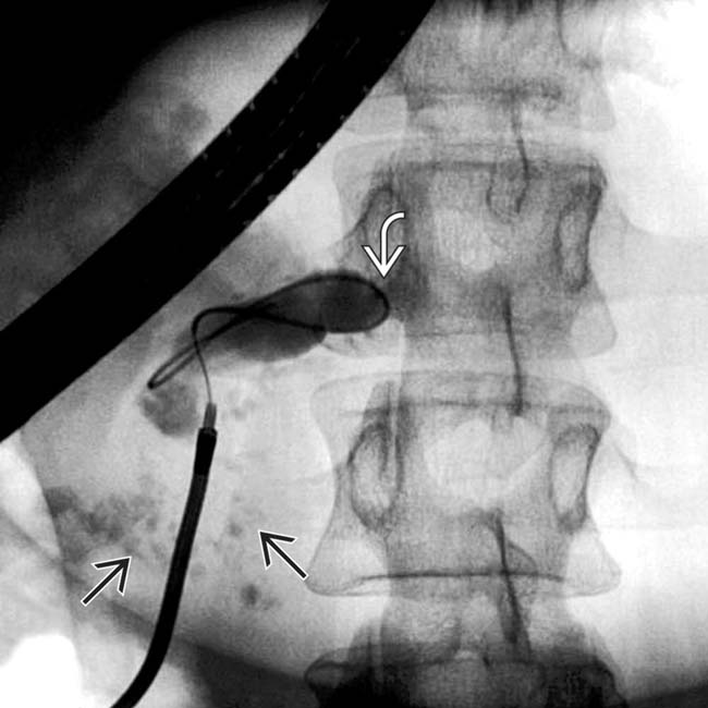
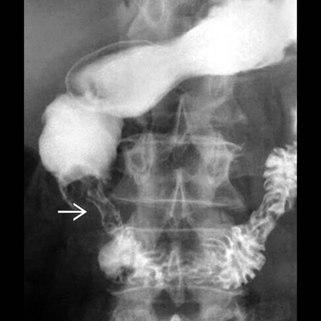
in the head of the pancreas. Note the dilated duct with abrupt obstruction to retrograde filling, due to a stone or stricture in the main pancreatic duct image.
image
Coronal MRCP in the same patient shows a signal void at the site of the main duct calculus image and a dilated, irregular duct upstream that could not be shown by the ERCP.
image
Axial T2WI shows an irregularly dilated main pancreatic duct image 
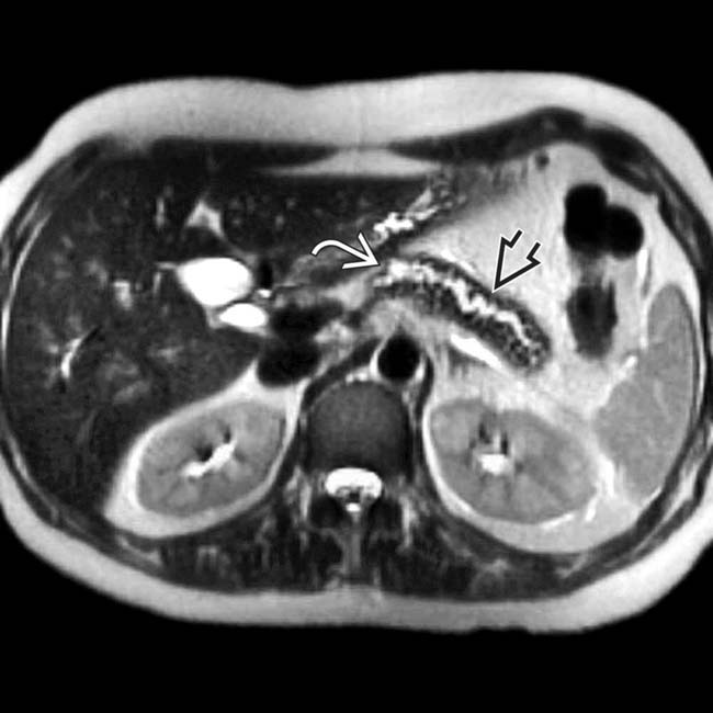
associated with glandular atrophy in the body-tail segments of the pancreas image.
image
Coronal MRCP MIP reconstruction after administration of secretin demonstrates a dilated, irregular pancreatic duct with subtle dilatation of several side branches. Fluid is seen reaching the transverse duodenum, compatible with grade III exocrine function.
image
Axial CECT demonstrates several calcifications image in the pancreatic head, virtually diagnostic of chronic pancreatitis, as well as a focal cyst image adjacent to the pancreas representing a pseudocyst.
image
Axial NECT demonstrates an atrophic pancreas with extensive calcifications, a virtually diagnostic appearance for chronic pancreatitis.
image
Axial CECT shows extensive calcifications in the pancreatic head.
image
Coronal MRCP MIP reconstruction demonstrates classic imaging findings of chronic pancreatitis, with a diffusely dilated main pancreatic duct and dilatation of many side branches.

Share this: