103
Chronic cutaneous lupus
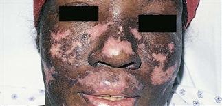
The scarred plaques of chronic cutaneous lupus have peripheral hyperpigmentation and central violaceous pink scarring. The lip is also involved.
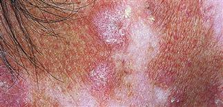
Typical lesion of cutaneous lupus, showing central hypopigmented scarring and a peripheral rim of pink scaling papules. This might be confused for a skin cancer or actinic keratosis.
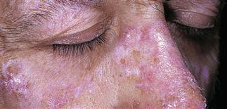
Scaling pink plaques, some with peripheral hyperpigmentation, scattered in a sun-exposed distribution on the face.
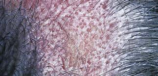
Lupus in the scalp, demonstrating alopecia. This lesion shows erythema, follicular prominence, and erosion. Discoid lesions in the scalp are a cause of scarring alopecia.
DESCRIPTION
Most common form of cutaneous lupus erythematosus. Lesions may be localized or widespread, and consist of scaling erythematous papules and plaques, often with central atrophy and scarring. Chronic cutaneous lupus lesions are not always discoid; this term should no longer be used.
HISTORY
• Chronic lupus erythematosus more common in women. • Perhaps more common in people with an African-American background. • Peak incidence in the fourth decade. • Trauma and ultraviolet light may initiate and exacerbate lesions. • Photo sensitivity is present in 50% of patients with discoid lupus erythematosus. • Lower incidence of systemic disease; 1–5% of cases progress to systemic lupus erythematosus. Patients with non-localized, widespread disease are at greater risk of advancing to systemic disease. • Scarring alopecia is permanent.
PHYSICAL FINDINGS
• Lesions may occur on any body surface, but scalp, face, and ears are most common areas. • Begins asymptomatically; there are well-defined, elevated, red to violaceous, 1- to 2-cm, flat-topped plaques with firmly adherent scaling. • Follicular plugs are prominent; peeling the scale reveals an undersurface that looks like a carpet penetrated by several carpet tacks. • Epidermal atrophy gives the surface either a smooth white or a wrinkled appearance. • Lesions may be hypertrophic or verrucous; they may involve palms and soles or the oral mucosa. • Lesions endure for months; they either resolve spontaneously or progress with further atrophy, ultimately forming smooth, white, or hyperpigmented depressed scars with telangiectasia and scarring alopecia. • Scalp disease begins with erythema, scaling, and follicular plugging. Hair follicles are destroyed, resulting in irreversible, scarring alopecia. Hair loss is haphazard in distribution. • Skin biopsy is helpful in making this diagnosis.
TREATMENT
• Treatment options include group I–III topical steroids, intralesional steroids, antimalarials (e.g. hydroxychloroquine 200 mg b.i.d.), dapsone, or oral corticosteroids. • Reasonable to start with a twice-daily application of a topical steroid and assess result after 6 weeks. If this fails, hydroxychloroquine could be considered. • Treatment options for difficult cases are methotrexate, azathioprine, thalidomide, or isotretinoin. • Sunscreens are an essential aspect of therapy. A broad-spectrum, water-resistant sunscreen should be applied daily. • Some patients may respond to tacrolimus ointment (Protopic).


