Choroidal Nevus
Clinical Features:
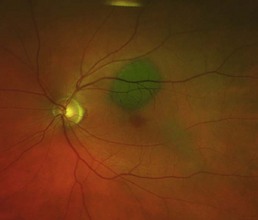
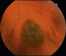
They are typically darkly pigmented, small, flat, and with well-defined borders (Fig. 21.1.1). Overlying drusen, present in Bruch’s membrane, are a common finding. Some nevi may have slight elevation (Fig. 21.1.2). They can occur throughout the fundus but are usually seen in the posterior pole. Accumulation of subretinal fluid, minimal growth over time, and alterations in pigmentation can occur in the absence of malignant transformation.
OCT Features:
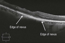
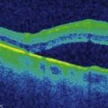
There is loss of the features of the choroicapillaris in the area of the nevus, which appears as a homogeneous well-defined area of hyper-reflectivity below the RPE (Figs 21.1.3 and 21.1.4). The overlying retinal layers are undisturbed. Enhanced depth imaging techniques can help to visualize the more posterior extent of a choroidal nevus (Fig. 21.1.5).
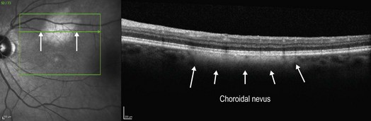
Figure 21.1.3 OCT (corresponding to Figure 21.1.1) shows a homogeneous, well-defined area of hyper-reflectivity below the RPE that obscures the features of the choriocapillaris in this region, corresponding to the choroidal nevus (arrows).
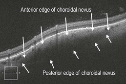
Figure 21.1.4 OCT (corresponding to Figure 21.1.2) shows a homogeneous well-defined area of hyper-reflectivity below the RPE that obscures the features of the underlying choriocapillaris centrally, but blends with the choriocapillaris on the edges. The overlying retina has a mild dome-shaped elevation due to the height of the lesion.