121
Chondrodermatitis nodularis helicis
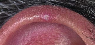
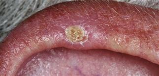
The primary lesion is a firm, tender, red to pink papule of 2–4 mm with a central keratotic punctum. It is most often found on the upper helix.
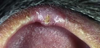
The central scale lacks the keratinous plug of keratoacanthoma. Removal of the scale reveals a small central erosion.
During the active stage, the base may become red and swollen; pain is constant.
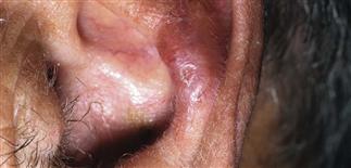
Chondrodermatitis nodularis helicis usually occurs in those over 40; the incidence increases with age. Lesions are occasionally found on the antihelix.
DESCRIPTION
An inflammatory process of the cartilage of the ear. It is an exquisitely tender papule on the most lateral edge of the helix or antihelix. Men are affected more often than women. The helix is involved more commonly in men, and the antihelix in women.
HISTORY
• Most patients are in the habit of sleeping on the affected side. Pressure from resting on a pillow causes pain, forcing the patient to alter sleeping position and affecting the ability to sleep comfortably. • The etiology is unclear. Related to focal dermal necrosis due to repetitive trauma. Over many years, dermal injury may result from actinic damage, physical pressure, or a combination of both. The vascular supply to this tissue is decreased, there is an inflammatory response, and damage is slow to heal. Inflammation and granulation tissue reflect attempts at healing the damaged collagen. • Without treatment, the lesions persist indefinitely. • Recurrences are common, even after aggressive therapy.
PHYSICAL FINDINGS
• The primary lesion is a firm, tender, red to pink, 2- to 4-mm papule with a central keratotic punctum. The punctum has firm, adherent crust or scale, resembling a small cutaneous horn. The surrounding skin shows scale, actinic damage with atrophy and telangiectasia. • Occasionally, there is more than one lesion. • The universal symptom is pain described as stabbing and sharp. • This condition is classically found on the most prominent and lateral portion of the auricle. • The major diagnostic consideration is squamous cell carcinoma, which tends to be more necrotic and less tender. A skin biopsy should be performed, which is diagnostic. A biopsy also rules out squamous cell carcinoma.
TREATMENT
• Any therapy must include efforts to relieve pressure and trauma on the affected area to allow healing. Patients who are able to sleep on their back should be encouraged to do so. Pillows should be positioned to minimize pressure on the ear. • Topical therapy is rarely successful. • Intralesional steroids can be effective in a minority of cases. Patients should expect some residual discomfort after injection. • Surgical removal of the lesion along with the inflamed cartilage can be curative. A shave excision is directed at removing all the surrounding inflamed tissue and cartilage. Curettage and light electrodesiccation of the base is performed, and the wound is allowed to heal by secondary intention. Definitive therapy involves surgical resection of the involved portion of the pinna. • Recurrences are common after any of the above therapies.


