16. Choanal Atresia
Definition
Choanal atresia is the congenital absence or closure of the paired openings between the nasal cavity and the nasopharynx. Because this obstruction is complete, the neonate with choanal atresia becomes an obligatory nose breather. The condition can cause death by asphyxiation in the newborn.
Incidence
The average incidence of choanal atresia is 0.82:10,000. Unilateral involvement occurs more frequently than the bilateral form by a ratio of about 2:1, with the right side being affected more often than the left. This condition affects females more frequently than males.
Etiology
During the normal uterine developmental process, the nasal cavities extend posteriorly under the influence of the posteriorly directed fusion of the palatal processes. The membrane thins, separating the nasal cavities from the oral cavity. The two-layer membrane that consists of nasal and oral epithelia ruptures and forms the choanae. This occurs by the 38th developmental day. If this rupture fails to occur, choanal atresia is the result. The “preliminary” choanae are not in the same location as the definitive choanae, which are eventually located even more posteriorly.
Signs and Symptoms
• Alleviation of respiratory distress by crying
• Arched hard palate
• Bilateral rhinorrhea
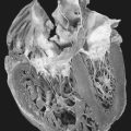
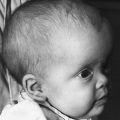
• Components of CHARGE (see box)
• Cyclical cyanosis
• Medialized lateral nasal well
• Narrow nasopharynx
• Severe airway obstruction
• Unilateral rhinorrhea
• Widened vomer
Medical Management
Choanal atresia is treated chiefly by surgical correction of the congenital anomaly. Bony involvement is present in 90% of patients. Bilateral atresia is generally corrected in the neonatal period. The surgical procedure is performed via a transnasal approach using either the CO 2 or Nd:YAG (neodymium:yttrium-aluminum-garnet) laser. Stents are placed in the newly created nasal passages for 3 to 5 weeks to maintain patency and improve continued postoperative passage patency. For the patient with unilateral atresia, definitive diagnosis may not occur for several years. Surgical correction should not be undertaken at any time during childhood.
Complications
• Aspiration
• Neonatal demise (from bilateral atresia)
• Respiratory arrest
Anesthesia Implications
Age-appropriate implications and concerns are foremost for infants and very young children scheduled for any surgical procedure. Corrective surgery for choanal atresia is no different.
Airway management may be difficult. Infants with CHARGE association should have their cardiac function thoroughly evaluated preoperatively.
CHARGE Association
Coloboma of the iris, choroid, and/or microphthalmia
Heart defect
Atresia of the choanae
Retarded growth and development
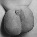
Genitourinary abnormalities (cryptorchidism, microphallus, hydronephrosis)
Ear defects associated with deafness
Co-morbidities or other associated abnormalities must be carefully considered and evaluated preoperatively.
The airway should be secured using an oral RAE tube after either inhalational or intravenous induction, whichever is more age appropriate.
Craniosynostosis Malformations
The patient should be extubated while as awake as possible with intact protective reflexes robust. However, if the surgical procedure has been prolonged, there may be significant airway edema and/or hemodynamic instability. In that case, the patient should remain intubated and sedated to allow for resolution of any such concerns before extubating and increasing the potential for catastrophic airway compromise.

