Chapter 1 Childhood Resuscitation
3 What are the common causes of cardiopulmonary arrest in children?
Common causes of cardiopulmonary arrest in children are numerous, but most fit into the classifications of respiratory, infectious, cardiovascular, traumatic, or central nervous system (CNS) diseases (Table 1-1). Respiratory diseases and SIDS together consistently account for one-third to two-thirds of all pediatric cardiopulmonary arrests in published series.
Table 1-1 Common Causes of Cardiopulmonary Arrest in Children
| Respiratory | Central Nervous System |
| Pneumonia | Seizures, or complications thereof |
| Near drowning | Hydrocephalus, or shunt malfunction |
| Smoke inhalation | Tumor |
| Aspiration and obstruction | Meningitis |
| Apnea | Hemorrhage |
| Suffocation | Other |
| Bronchiolitis | Trauma |
| Cardiovascular | Sudden infant death syndrome |
| Congenital heart disease | Anaphylaxis |
| Congestive heart failure | Gastrointestinal hemorrhage |
| Pericarditis | Poisoning |
| Myocarditis | |
| Arrhythmia | |
| Septic shock |
8 After establishing a clear chain of command and assigning specific duties to all members of the resuscitation team, what should the order of priorities be?
9 What is the recommended way to establish a patent airway?
 The first attempt to establish airway patency should be through proper airway positioning. Often, this alone will be effective. Since most airway obstruction is due to the effect of gravity on the mandibular block of soft tissues, it can be relieved by either a head tilt–chin lift or jaw-thrust maneuver.


10 What is the recommended way to deliver supplemental oxygen to a child?
Supplemental oxygen can be delivered to a child by a variety of different means. For the sickest patient, oxygen should be delivered in the highest concentration and by the most direct method possible. Children who demonstrate spontaneous breathing might require less invasive means of administration of supplemental oxygen. Table 1-2 lists some different methods of oxygen delivery with their associated delivery capabilities.
Table 1-2 Methods of Oxygen Delivery and Their Delivery Capabilities
| Nasal cannula: 30–40% oxygen |
| Simple masks: 30–60% oxygen |
| Partial rebreather masks: 50–60% oxygen |
| Oxygen tents: 30–50% oxygen |
| Oxygen hoods: 80–90% oxygen |
| Nonrebreather masks: ~100% oxygen |
11 Which children require intubation?
 Inadequate central nervous system control of ventilation
 Functional or anatomic airway obstruction

 Loss of protective airway reflexes
 Excessive work of breathing, which might lead to fatigue and respiratory insufficiency
 Need for high airway pressures to maintain effective alveolar gas exchange
 Need for mechanical ventilatory support
 Potential occurrence of any of the above during patient transport
12 When selecting an endotracheal tube, what sizing guidelines are suggested?

KEY POINTS: HOW TO DETERMINE THE PROPER PLACEMENT OF THE ETT
1 Check to see that the tube is inserted at a depth that is three times the internal diameter of the ETT (from the point of the patient’s central incisors).
2 Observe for symmetric chest expansion.
3 Auscultate for symmetric breath sounds.
4 Look for distention of the abdomen, indicating misplacement of the tube.
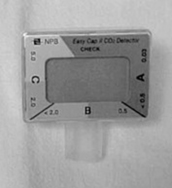
5 Measure end-tidal carbon dioxide using a colorimetric detector. In infants and children with a perfusing rhythm, a purple color on the device indicates a problem, whereas a yellow color implies that the tube is in the trachea.
13 How can I determine if ETT placement is appropriate?
Proper depth for ETT insertion from the point of the patient’s central incisors can be estimated to be three times the internal diameter of the ETT. Measurement of end-tidal carbon dioxide using a colorimetric detector, observation for symmetric chest expansion, and auscultation for symmetric breath sounds can help to ensure proper placement. Confirmation of placement is probably best determined with a chest radiograph. Prior to an x-ray, the colorimetric detector offers a rapid bedside determination to detect CO2 to confirm endotracheal tube placement (Fig. 1-1).
14 What are the best methods to assess a child’s circulatory status?
Assessment of a child’s circulatory status should always include appraisal of:


16 What are the golden rules of vascular access?
20 What role does drug therapy play in pediatric resuscitation?
 Epinephrine (to increase heart rate, myocardial contractility, and systemic vascular resistance)
 Atropine (to increase heart rate in nonneonates)
 Dextrose (to increase glucose)
 Sodium bicarbonate (to increase pH)
 Amiodarone or procainamide (to reverse ventricular arrhythmias)
 Naloxone (to reverse the effects of narcotics)
 Adenosine (to reverse supraventricular tachycardia)
 Dopamine (to increase vasoconstriction and blood pressure)


21 What are the new recommendations regarding epinephrine administration during pediatric resuscitation?
PALS recommendations for pulseless arrest (PES, asystole)
 If the first asystole-countering dose is intravascularly (IV or IO route) administered: give as a standard dose (0.01 mg/kg). This can be delivered as 0.1 mL/kg of a 1:10,000 solution of epinephrine. Vasopressin is used in place of epinephrine for the first or second dose in adult resuscitations but is considered Class Indeterminate (not enough evidence to recommend for or against) in pediatric arrests.


PALS recommendations for bradycardia


22 Which resuscitation drugs are effective when given via an endotracheal tube?
KEY POINTS: DRUGS THAT CAN BE GIVEN VIA THE ENDOTRACHEAL ROUTE
23 Are there minimum dosing requirements for any resuscitation drugs?
 Atropine (usual dose, 0.02 mg/kg) has a minimum dosing requirement for effective reversal of bradycardia. It appears that at doses lower than 0.1 mg, atropine exerts an effect that might actually worsen bradycardia. Thus, if its use is considered for reversal of bradycardia in a child who weighs less than 5 kg, a minimum of 0.1 mg should be administered.


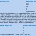
27 Is there an easy method to calculate mixtures of constant infusions of drugs?
Several methods are used. Here is one easy method:
 For constant infusion of drugs (epinephrine, isoproterenol) beginning at 0.1 μg/kg/min: 0.6 times the weight in kg equals the number of milligrams of drug to add to enough water to make a total of 100 mL of solution. The resultant solution is then infused at a rate of 1 mL per hour, delivering 0.1 μg/kg/min.