Chapter 8 Chest Pain
4 Which diagnoses are most common in children who present to an ED with chest pain?
In many studies, up to 45% of cases of chest pain in children are labeled “idiopathic.” That is, after a careful history and physical examination, the etiology is still uncertain. When a diagnosis can be found, musculoskeletal injury is most common. Active children frequently strain chest wall muscles while carrying heavy books, exercising, or engaging in rough play. Many other children suffer chest pain from a direct blow to the chest that results in a mild contusion or, in rare cases, a rib fracture. Costochondritis accounts for about 10–20% of cases of chest pain. This musculoskeletal disorder produces tenderness over the costochondral junctions and is often bilateral. It is exaggerated by physical activity or breathing. Musculoskeletal pain is often reproducible by palpation of the chest wall or moving the arms and chest through a variety of positions. Table 8-1 lists the most common etiologies for pediatric chest pain.
| Idiopathic Musculoskeletal |
| Chest wall strain |
| Costochondritis |
| Direct trauma |
| Respiratory conditions |
| Asthma |
| Cough |
| Pneumonia |
| Gastrointestinal problems |
| Esophagitis |
| Esophageal foreign body |
| Psychogenic (stress related) |
| Cardiac pathology |
| Myocarditis |
Selbst SM: Chest pain in children—consultation with the specialist. Pediatr Rev 18:169–173, 1997.
8 What is “slipping rib syndrome”?
Mooney DP, Shorter NA: Slipping rib syndrome in childhood. J Pediatr Surg 32:1081–1082, 1997.
11 When should a pneumothorax be suspected in a child with chest pain?
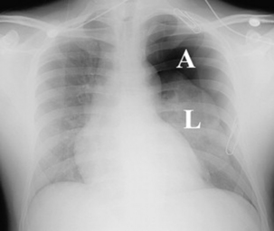
Suspect a pneumothorax if a child develops acute onset of sharp chest pain associated with some degree of respiratory distress. The pain is usually worsened by inspiration and may radiate to the shoulder, neck, or even the abdomen. Children with this condition do not have long-standing pain and almost all present for care within 48 hours of developing the pneumothorax. The patient will usually have dyspnea, tachycardia, and, perhaps, decreased breath sounds on the affected side, or even cyanosis. However, these signs and symptoms depend on the size of the pneumothorax and whether it is under tension (Fig. 8-1). A small pneumothorax may produce minimal findings on examination.
14 Which children with chest pain deserve an evaluation with an electrocardiogram and/or chest radiograph?
15 Which children with chest pain do not need extensive evaluation with laboratory studies in the ED?
18 Which cardiac conditions can cause chest pain in children?
 Arrhythmia: Supraventricular tachycardia, ventricular tachycardia
 Infection: Myocarditis, pericarditis
 Structural abnormalities: Hypertrophic cardiomyopathy, aortic valve stenosis, anomalous coronary arteries
 Coronary artery disease (CAD; ischemia or infarction): Kawasaki disease, long-standing diabetes mellitus, familial hypercholesterolemia or lipidemia
21 When should I be concerned about a cardiac infection as the cause of chest pain?
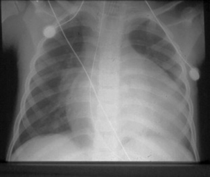
Children with infections such as myocarditis or pericarditis can present with chest pain and usually have fever. Those with pericarditis may report a sharp, stabbing, midsternal pain that is somewhat relieved when the patient sits up and leans forward. Distant heart sounds, neck vein distention, and a friction rub may be found. Viral myocarditis is more common and usually presents with low-grade fever and dull substernal chest pain. There is often respiratory distress as the infection progresses, and there may be muffled heart sounds or a gallop rhythm heard. Tachycardia out of proportion to the degree of fever is characteristic. Tachycardia and hypotension may be worse with standing, and this may not resolve after fluid resuscitation. A chest radiograph often reveals cardiomegaly (Fig. 8-2).
22 Which cardiac conditions can lead to ischemia and chest pain?
27 What etiology should be considered in an afebrile, previously well toddler, with no injury, who reports sudden midsternal chest pain?
30 Name a cutaneous condition that is associated with chest pain.
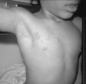
Herpes zoster infection (Fig. 8-3). Shingles is often associated with distressing chest pain when the lesions involve a chest wall dermatome. The chest pain can sometimes precede the vesicular rash by several days.
35 Chest pain associated with a “crunching” sound on chest examination is found with which condition?
Kanegaye JT: An adolescent football player with chest pain. Clin Pediatr 42:471–474, 2003.


