142
Cherry angioma
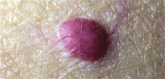
Early lesions are bright-red, smooth, dome-shaped papules.
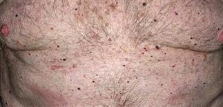
Early lesions occur mostly on the trunk.
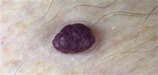
Older lesions develop gray strands of fibrosis, producing a multilobulated appearance.
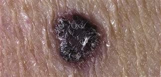
Cherry angiomas may mimic pigmented nevi and melanoma. Magnification or dermoscopy can be helpful to distinguish the two.
DESCRIPTION
Benign vascular neoplasm found in nearly all people older than 30 years.
HISTORY
• Lesions appear gradually in adulthood and are asymptomatic. • Multiple eruptive cherry angiomas have been associated with bromide exposure, sulfur mustard gas, and the glycol ether solvent 2-butoxyethanol. • The sudden appearance of multiple lesions may warrant a search for occult malignancy, especially for small tumors capable of hormone production (those in the pancreas, small bowel, and respiratory system). • Undisturbed cherry angiomas persist indefinitely. • Superficial trauma may produce bleeding. • Isolated reports of patients with hundreds of cherry angiomas arising in association with pregnancy, and also in patients with elevated prolactin levels, suggest that hormonal factors may play a role.
PHYSICAL FINDINGS
• A few to hundreds of discrete, 0.5- to 5.0-mm, smooth, dome-shaped to polypoid papules occur mostly on the trunk but can be found on the head, neck, and extremities. • Early smaller lesions are cherry red, deeper larger lesions maroon.
TREATMENT
Ablation with electrocautery, laser surgery, cryosurgery, or by simple scissor excision carries slight risk of scarring and dyspigmentation.