56 Charcot–Marie–Tooth disease (peroneal muscular atrophy)
Salient features
Examination
• Wasting of muscles of calves and thighs that stops abruptly, usually in the lower third of the thigh, and is described as ‘stork’ or ‘spindle’ legs, ‘fat bottle’ calves and ‘inverted champagne bottles’ (Fig. 56.1)
• Pes cavus (high arched foot) or pes planus (flat foot), clawing of toes, contractures of the Achilles tendon
• Weakness of dorsiflexion, foot drop
• Absent ankle jerks, plantars are downgoing or equivocal
• Mild sensory impairment or no sensory loss. Some patients have decreased responses to pain in the stocking distribution.
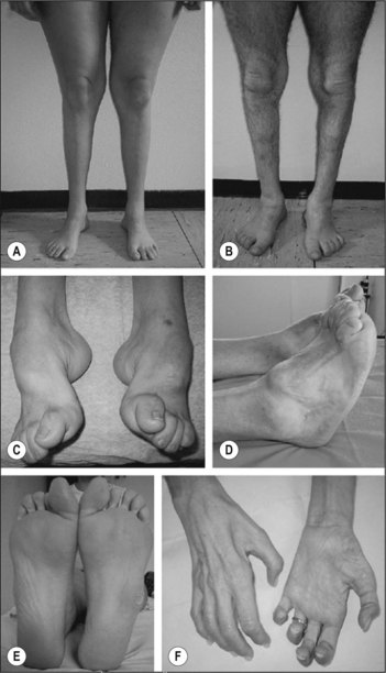
• Feel for lateral popliteal nerve thickening (seen in some cases only)
Note: There are two distinctive clinical features of this disease:
1 The muscular atrophy begins in the distal portions of the affected muscles in the lower and upper limbs, unlike the global atrophy of motor neuron disease or muscular dystrophy. The atrophy then creeps upwards, involving all muscles.
2 Second, the degree of disability is minimal in spite of marked deformity.
Advanced-level questions
What is the mode of inheritance?
• Each type can be inherited in a dominant, recessive or X-linked fashion. There are also autosomal dominant intermediate forms of CMT that can have features of both axonal and demyelinating neuropathies. Mutations in 31 known genes and additional unidentified loci can produce CMT:
• Current evidence-based clinical guidelines for distal symmetric polyneuropathy recommend genetic testing consisting of screening for common mutations, including the CMT1A duplication copy-number variant and point mutations of the X-linked GJB1.
• Whole genome sequencing is emerging to be the diagnostic tool in this condition.
Mention other hereditary neuropathies
• Roussy–Lévy syndrome (where features of progressive muscular atrophy may be combined with tremor and ataxia)
• Refsum’s disease (phytanic acid accumulates in the central and peripheral nervous systems)
• Fabry’s disease (where there is a deficiency of α-galactosidase)
• Bassen–Kornzweig disease (abetalipoproteinaemia, absence of LDL, and vitamin E deficiency)
• Metachromatic leukodystrophy (where galactosyl sulfatide accumulates in the central and peripheral nervous systems).


