Challenge

Published on 03/05/2015 by admin

Filed under Radiology

Last modified 22/04/2025

Print this page

rate 1 star rate 2 star rate 3 star rate 4 star rate 5 star
Your rating: none, Average: 0 (0 votes)

This article have been viewed 1616 times

Chapter 3 Challenge

Comment

The HPEs are a spectrum of disorders characterized by hypoplasia of the rostral end of the neural tube and the premaxillary segment of the face (lack of forebrain induction). HPE is characterized by failure of cleavage of the embryonic prosencephalon, which is normally complete by embryonic day 35. With this comes partial to complete failure of separation of the telencephalon and diencephalon into the right and left cerebral hemispheres and basal ganglia or thalami, respectively. Because the optic vesicles and olfactory bulbs evaginate from the prosencephalon, visual disturbances and incomplete formation of the olfactory system are frequently present. Hypoplasia of the premaxillary segment results in facial anomalies, including cleft lip and palate; abnormalities of the orbit (cyclopia, hypotelorism); and forehead proboscis.

Holoprosencephaly may be divided into subtypes: alobar (the most severe form), semilobar, lobar (the mildest form of the major subtypes), and middle interhemispheric variant. There is no clear distinction between subtypes. In alobar HPE, the falx, interhemispheric fissure, and septum pellucidum are absent. There is failure of separation of the cerebrum and the ventricular system, resulting in a monoventricle contiguous with a dorsal cyst. In semilobar HPE, the interhemispheric fissure and falx cerebri are usually formed posteriorly and absent anteriorly. In lobar HPE, the interhemispheric fissure and falx anteriorly are hypoplastic. Often the posterior corpus callosum or splenium is formed, as in this case (arrow). The third ventricle is usually well formed. In semilobar and lobar HPE, the ventricular system shows variable degrees of development. The middle interhemispheric variant differs from classic HPE in that the posterior frontal and parietal lobes are most significantly affected rather than the basal forebrain. The anterior frontal lobes and occipital lobes are separated, and the genu and splenium of the corpus callosum are formed, but the callosal body is absent. The hypothalamus and lentiform nuclei appear separated, but the caudate nuclei and thalami are incompletely separated in many cases. Heterotopias and cortical dysplasia are common associated anomalies of HPE, as seen in this case, in the left greater than right medial frontal lobes.

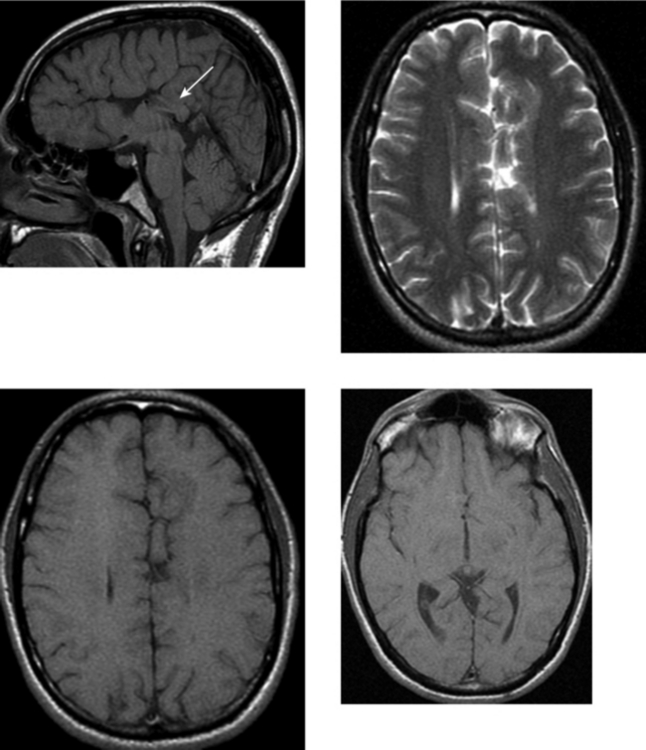
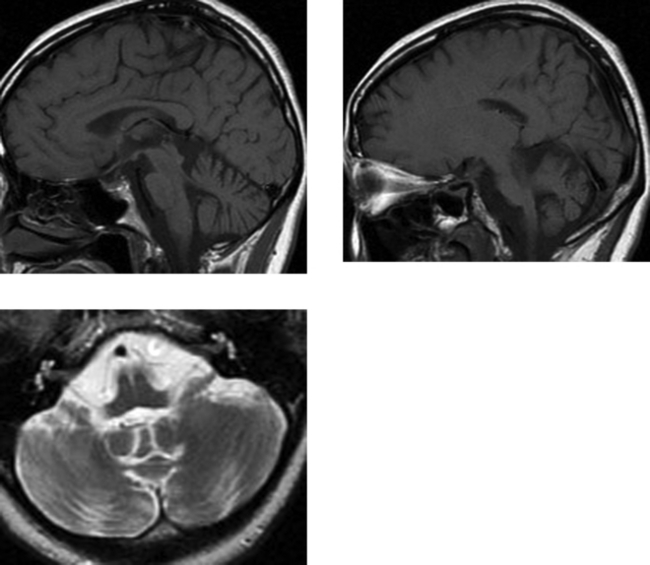
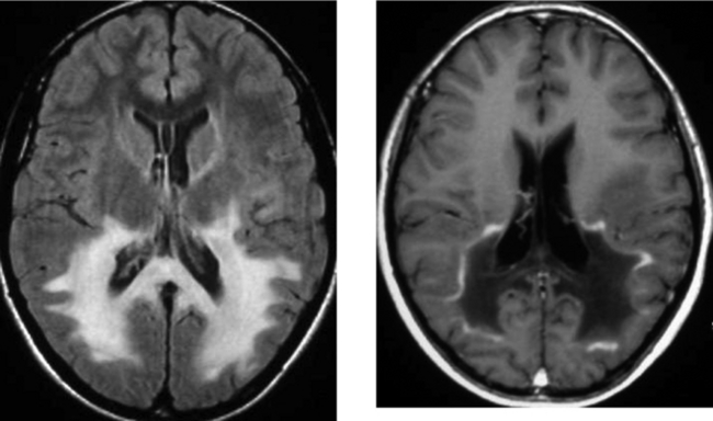
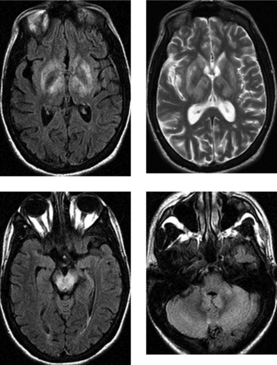
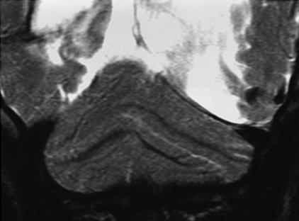
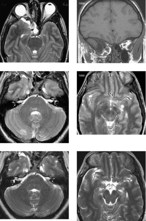
CASE 152 Olivopontocerebellar Degeneration

CASE 153 Band Heterotopia—Pachygyria

Comment

Heterotopias are migrational abnormalities in which normal neurons occur in abnormal locations as a result of failure of migration along the radial glial fibers from the germinal region to the cortex. Three well-recognized types of heterotopia include focal, subependymal, and diffuse (laminar, band). Subcortical heterotopia is a newer term that refers to a specific entity in which neurons are abnormally located predominantly in the subcortical white matter. The cortical dysplasias are different from the heterotopias in that they do not represent a failure of normal migration, but rather a failure in the development of a normal six-layered cortex after migration from the germinal matrix to the cortical region.

Diffuse or band heterotopia refers to a layer of gray matter whose migration has arrested such that it is localized between the subcortical white matter laterally and the deep white matter medially. The inner and outer margins of the heterotopic neurons are well demarcated. Therefore, the band of gray matter is separated from the overlying cortex by a mantle of subcortical white matter. Band heterotopias are frequently associated with overlying cortical dysplasias (pachygyria, polymicrogyria), as in this case. The severity of the cortical dysplasia is directly related to the severity of the band heterotopia: the thicker the band, the more severe the overlying dysplasia will be. Similarly, patients with band heterotopia tend to have more pronounced clinical symptoms. In addition to seizures, which may have an age of onset ranging from infancy to young adulthood, many children have moderate to severe developmental delay.

Hemimegalencephaly refers to hamartomatous overgrowth of a cerebral hemisphere. Migrational abnormalities are usually present within the affected hemisphere, but they may also be present on the contralateral side. On imaging, there is usually enlargement of the affected cerebral hemisphere and the ipsilateral lateral ventricle, as well as associated cortical dysplasias.

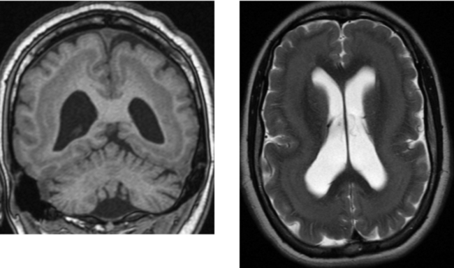
CASE 154 Huntington’s Disease

Comment

A variety of disease processes affect the extrapyramidal nuclei (basal ganglia, thalami) as well as the nuclei in the brainstem. These conditions are most commonly degenerative or metabolic, and many are inherited. Among the neurodegenerative processes that can affect the deep gray matter are Huntington’s disease, Wilson’s disease, and Hallervorden-Spatz syndrome. Toxic exposures may also result in abnormalities of the deep gray matter. Lesions in these structures typically result in movement disorders that can occur in isolation or in combination and include the following subtypes: abnormalities in muscle tone, involuntary movements, abnormal postural reflexes, and the inability to carry out voluntary movements. Toxic exposures that affect the basal ganglia include carbon monoxide, cyanide, hydrogen sulfide, ethylene glycol, and toluene. Although there is wide variation in the deep gray matter structures involved, many diseases have characteristic involvement of specific structures.

Huntington’s disease is inherited in an autosomal dominant manner that results in progressive degeneration of brain cells. The gene responsible for the disease is located on chromosome 4. Clinical manifestations typically include involuntary movement (choreoathetosis), rigidity, dementia, and emotional instability. Cognitive deficits are believed to be due to abnormal connectivity between the deep gray matter and the cortex. The disease typically presents in the fourth or fifth decade of life. The disease is progressive, with death occurring 15 to 20 years after its onset. On imaging, as in this case, Huntington’s disease is characterized by atrophy of the caudate nuclei, which results in ballooning of the frontal horns of the lateral ventricles (“boxcar” ventricles). There may also be involvement of the putamen, which can atrophy. MR imaging shows signal changes in these nuclei. These changes may be hyperintense on T2W images, as in this case, which is believed to be related to gliosis; other cases show T2W hypointensity, which is likely related to iron deposition. Other major imaging findings include cortical atrophy.

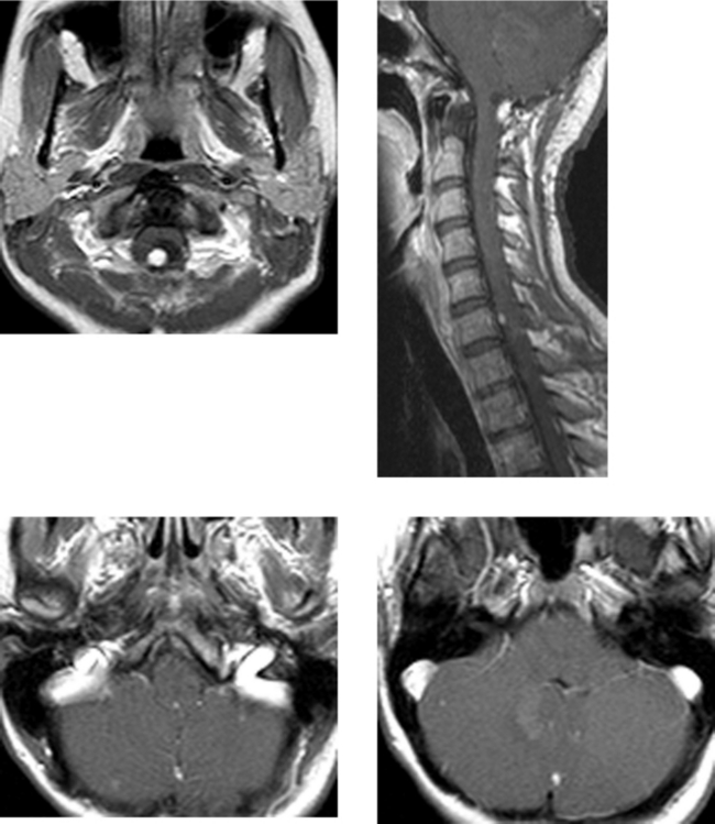
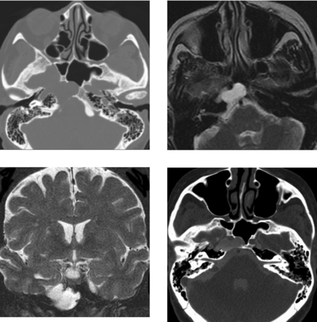
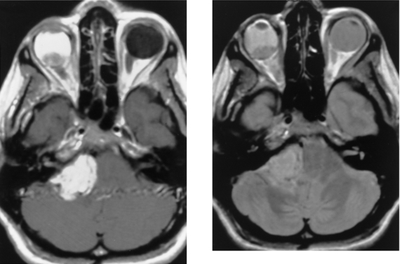
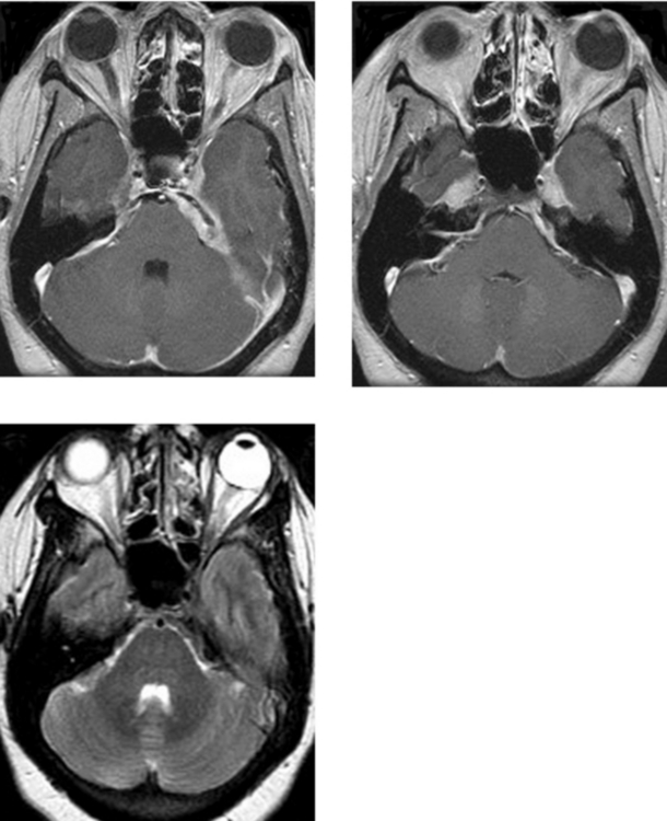
CASE 155 Meningocele and Pseudomeningocele of the Skull Base (Petrous Apex)

Comment

This case shows a well-demarcated, chronic-appearing lesion of the right petrous apex. There is expansion, smooth cortical bone thinning without bone destruction, and cortication medially, suggesting a long-standing process. The lesion is isointense to CSF on T2W images, and on the coronal T2W image, it is difficult to determine whether this lesion is separate from or an extension of Meckel’s cave into the osseous base of the skull. CT cisternography (instillation of contrast material into the thecal sac by lumbar puncture, followed by CT imaging after graceful tilting of the patient so that the contrast agent spreads through the subarachnoid spaces in the head) confirms that the petrous apex lesion communicates with the CSF spaces.

The differential diagnosis of a benign-appearing, expansile petrous apex mass includes cholesterol granuloma, mucocele, epidermoid, meningocele, and occasionally, an aneurysm of the internal carotid artery. Mucoceles are frequently unilocular and show peripheral enhancement. The signal characteristics of mucoceles will vary, depending on the protein concentration and viscosity. Epidermoid cysts may have a benign appearance, or they can have more concerning radiologic findings, such as bone erosion or destruction. They typically appear similar to CSF on T1W and T2W images and hyperintense on FLAIR imaging, and demonstrate restricted diffusion. Cholesterol granulomas, when large, may be multilocular. There is usually thinning of the cortex with large lesions without bone destruction. Cholesterol granulomas are often hyperintense on all pulse sequences. In particular, the marked hyperintensity on unenhanced T1W images distinguishes these from many other lesions, and the presence of hemorrhage–fluid levels within these lesions is highly characteristic.

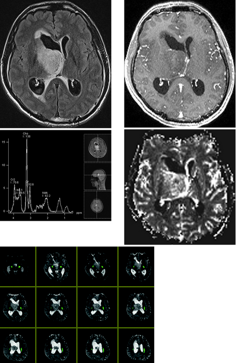
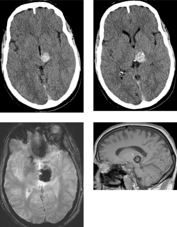
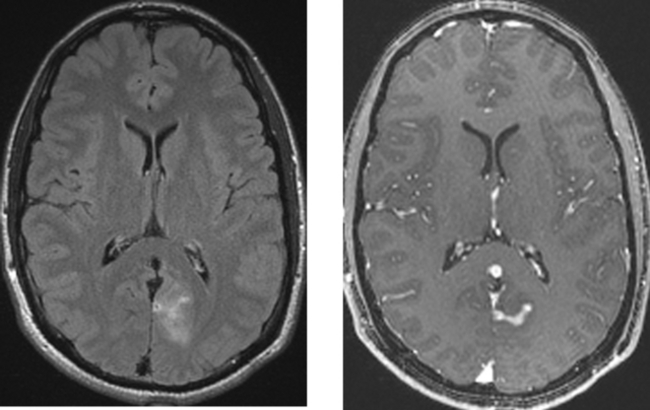
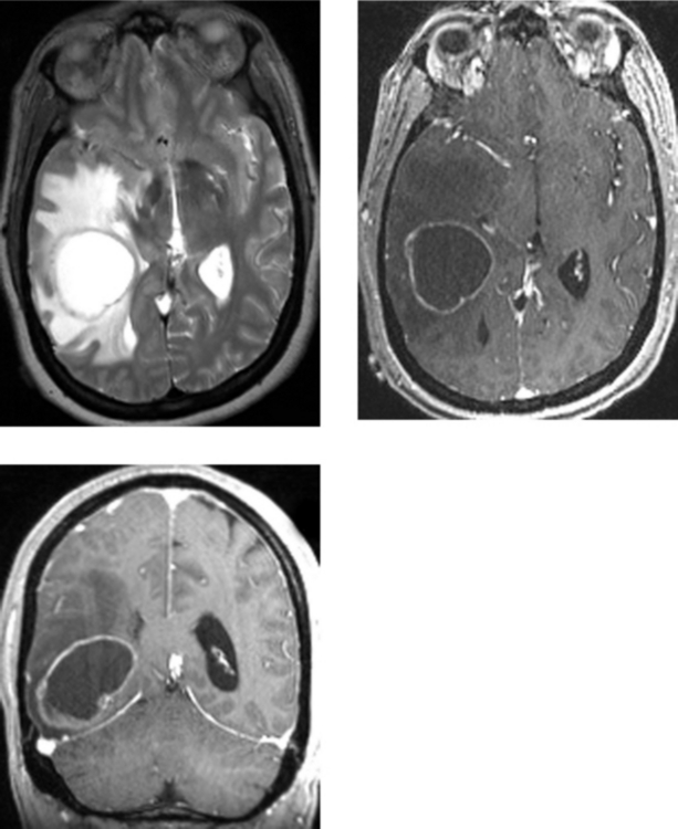
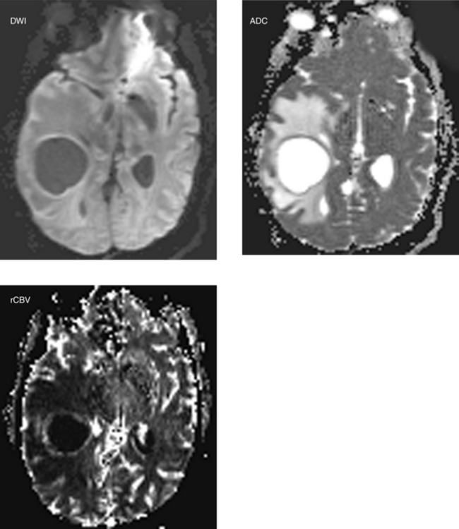
CASE 156 Right Thalamic Glioblastoma

Comment

This case illustrates a solid mass centered in the right thalamus, with an exophytic component extending into the ventricle. There is mass effect, with partial obstruction of the foramen of Monroe resulting in hydrocephalus and transependymal flow of CSF along the margin of the right lateral ventricle. Most GBMs enhance and usually demonstrate heterogeneity because of the presence of necrosis or hemorrhage. Margins of the enhancing component are usually ill defined, and there is irregular thick peripheral enhancement due to central necrosis. Enhancement may extend into the adjacent white matter. In a newly identified brain tumor in which biopsy is anticipated, regions of enhancement correlate with regions of solid tumor on pathologic examination. Therefore, contrast is useful in identifying areas for stereotactic biopsy. In addition, enhanced images may identify tumor spread to regions that otherwise would not be noticed on unenhanced images, such as the leptomeninges, subarachnoid space, or subependymal region along the ventricular margins.

In this less typical case, the GBM is centered in the deep gray matter (most GBMs occur in the cerebral lobes), has no significant central necrosis, and shows only mild central enhancement. The functional in vivo techniques of MR spectroscopy and MR perfusion imaging allow correlation of metabolic activity with the vascular properties, respectively, and provide further insight into the grade of the primary glial neoplasm. In this case, these techniques were helpful in characterizing the high-grade nature of this neoplasm. Specifically, proton spectroscopy shows an elevated choline:NAA ratio (>2), and perfusion imaging shows markedly elevated rCBV, indicative of a high-grade neoplasm. For the neurosurgeon planning stereotactic biopsy, location of eloquent areas of brain relative to the tumor is critical to reduce potential neurologic deficits. Diffusion tensor imaging tractography in this case clearly identifies the corticospinal tracts (green).

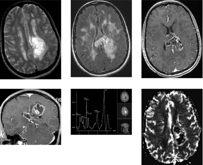
CASE 157 Multiple Sclerosis and Glioblastoma

Comment

“Tumefactive” multiple sclerosis, high-grade glioma (GBM), and occasionally an abscess can appear similar on imaging, particularly in the absence of a clinical history. Multiple sclerosis typically occurs in younger patients, and there are often additional clinical or imaging findings to suggest this diagnosis. On close questioning, patients often have neurologic symptoms that are spaced both in time and in location. Furthermore, MR imaging may demonstrate white matter lesions separate from the mass that are suggestive of multiple sclerosis.

Advanced MR imaging sequences may be of value. Perfusion imaging in this case shows markedly elevated regional cerebral blood volume in the enhancing rim of the necrotic mass, suggesting a high-grade glioma or GBM rather than demyelination. GBM was confirmed at surgical biopsy. Nonspecific spectroscopic findings in tumefactive multiple sclerosis include elevation of choline, lactate, and lipid peaks, and a decrease in the N-acetylaspartate peak. These spectroscopic characteristics reflect the histologic correlate of marked demyelination in the absence of significant inflammation. Gliomas also consistently show reductions in N-acetylaspartate and increases in phospholipids, reflecting the replacement of normal neuronal tissue with a proliferating cellular process. Increases in lactate are not uncommon as a result of tissue ischemia and necrosis, as in this case. Variable increases in lipid levels may be seen.

Elevation of glutamate or glutamine peaks favors tumefactive demyelination and is typically not seen in aggressive neoplasms (not present in this case). Serial proton MR spectroscopy can be a useful, noninvasive method of overcoming the diagnostic dilemma of differentiating glioma from acute tumefactive demyelination. Persistent elevation of choline and lactate levels favors a glioma. Normalization of the initial increases in phospholipids, lipid, and lactate peaks within 3 to 4 weeks, followed by persistent, marked reductions of the neuronal marker N-acetylaspartate, has been described over time in tumefactive demyelination.

CASE 158 Adrenoleukodystrophy

Comment

Adrenoleukodystrophy is an X-linked or autosomal recessive disorder that is related to a single enzyme deficiency (acyl coenzyme A synthetase) within intracellular peroxisomes. This enzyme is necessary for β oxidation in the breakdown of very long-chain fatty acids that accumulate in erythrocytes, plasma, and fibroblasts, as well as the CNS white matter and adrenal cortex. Boys typically present between the ages of 4 and 10 years. The clinical presentation may include behavioral disturbance, visual symptoms, hearing loss, seizures, and eventually spastic quadriparesis. Patients often present with adrenal insufficiency (Addison’s disease), which may occur before or after the development of neurologic symptoms.

As in other demyelinating and dysmyelinating disorders, MR is the imaging modality of choice for the detection of white matter disease, being far superior to CT. In adrenoleukodystrophy, the most common pattern of white matter disease is bilaterally symmetric abnormalities within the parietal and occipital white matter, extending across the splenium of the corpus callosum. The disease may continue to progress anteriorly to involve the frontal and temporal lobes. The region of active demyelination, usually along the anterior margin, may show contrast enhancement. Less typical presentations include predominantly frontal lobe involvement or holohemispheric involvement. Adrenoleukodystrophy also involves the cerebellum, spinal cord, and peripheral nervous system. Findings on MR imaging correlate well with neuropsychological measures.

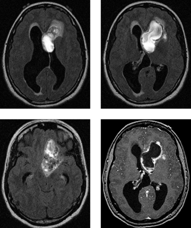
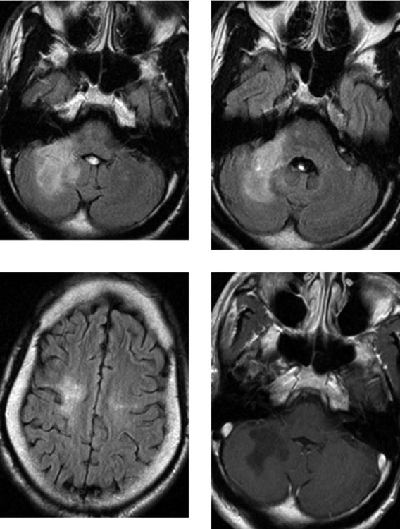
CASE 159 Medulloblastoma

Comment

Medulloblastomas account for up to one third of all pediatric posterior fossa tumors. They occur more commonly in boys than in girls (approximately 3:1) and arise from the superior medullary velum of the fourth ventricle from primitive neuroectoderm. In children, they are typically midline masses associated with the inferior vermis, but occasionally may present as a lateral cerebellar hemispheric mass, as in this case. Importantly, subarachnoid seeding of the leptomeninges is very common at presentation (reported in up to 30% of cases in some series); therefore, patients should have a screening contrast enhanced MR imaging study of the spine to exclude this type of spread.

On unenhanced CT, medulloblastomas are typically hyperdense relative to brain parenchyma because of their dense cellularity. They are demarcated masses, and calcification, cystic change, or hemorrhage may be present in up to 10% to 20% of lesions. On MR imaging, the signal characteristics of medulloblastomas vary considerably on T2W imaging, depending on the presence of hemorrhage and the degree of cellularity. Most medulloblastomas show avid but heterogeneous contrast enhancement. Typically, they efface the fourth ventricle and present with hydrocephalus.

Accurate preoperative diagnosis is important in pediatric cerebellar tumors because this may affect the surgical approach. Diffusion MR imaging allows assessment of microscopic water diffusion within tissues, and with neoplasms, this diffusion seems to be primarily based on cellularity. Increasing cellularity leads to increased signal intensity on diffusion imaging and hypointensity on corresponding apparent diffusion coefficient (ADC) maps. Studies have suggested that diffusion imaging and ADC values may be useful in helping to distinguish among histologic types of pediatric brain tumors. Juvenile pilocytic astrocytomas have shown high ADC values and ratios. In contrast, medulloblastomas that characteristically are cellular have shown restricted diffusion, as in this case, with high signal intensity on diffusion images (image 3) and hypointensity on ADC maps (image 4). The cellularity of ependymomas is between that of astrocytomas and that of medulloblastomas. Hence, pilocytic astrocytomas are most often hyperintense on ADC maps, medulloblastomas are hypointense, and ependymomas fall somewhere in between.

CASE 160 Middle Cerebral Artery Territory Stroke Mimicking a Bone Metastasis

Comment

The left lateral projection from the patient’s delayed bone scan shows increased radiotracer activity projecting over the temporal bone region (it is important to remember that an anteroposterior view is necessary to accurately localize the abnormality). This was the only abnormality on the patient’s bone scan. Corresponding enhanced MR imaging shows mild local mass effect in the superficial portion of the posterior left temporal lobe with cortical enhancement; the imaging findings are consistent with a stroke. In this patient with renal cell carcinoma, the bone scan was initially interpreted as showing metastatic disease. It is important to remember that any process that stimulates deposition of calcium within soft tissues, the solid organs, or areas of infarction may result in increased uptake of technetium-labeled radiopharmaceuticals. Similarly, in circumstances in which there is increased blood flow or luxury perfusion, there will also be increased radiotracer uptake. Infarctions in the brain may take up radionuclide, as can brain metastases from systemic cancers, which have a predilection to calcify (mucinous adenocarcinomas, including breast and gastrointestinal carcinomas; sarcomas; and in children, neuroblastomas). In a patient with a known systemic malignancy, an isolated region of increased radiotracer uptake in the cranium should be further assessed with additional imaging (plain films, CT, or MR imaging as needed) before it is assumed to represent metastatic disease. In this patient, MR imaging was obtained 1 week after the bone scan because of new-onset left upper extremity weakness. Images showed an acute infarct in the distal right middle cerebral artery territory (not shown) and, at the same time, confirmed a subacute to chronic infarct in the left middle cerebral artery territory. In addition, no calvarial lesion was identified on MR imaging.

CASE 161 Cytomegalovirus Meningitis and Ependymitis in a Patient with AIDS

Comment

Cytomegalovirus is present in the latent form in the majority of the American population. Reactivation usually results in a subclinical or mild flu-like syndrome. In immunocompromised patients, reactivation can result in disseminated infection, usually involving the respiratory and gastrointestinal tracts; however, rarely, it can infect the nervous system. In the CNS, CMV may cause meningoencephalitis and ependymitis. Symptoms may be acute or chronic, developing over months. Patients may have fever, altered mental status, and progressive cognitive decline. Patients may also present with cranial neuropathies (as in this case). CMV polymerase chain reaction in the CSF is sensitive and specific for the diagnosis of AIDS-related CMV infection of the CNS. However, conventional CSF findings and neuroimaging may not adequately assess the severity of CNS CMV disease, as demonstrated at autopsy.

Magnetic resonance imaging is the diagnostic study of choice in assessing immunocompromised patients suspected of having CNS infection. Imaging may show atrophy; high signal intensity in the periventricular white matter, typically not associated with significant mass effect; and retinitis (frequently seen in the AIDS population) in patients with CMV infection. Although patients with CNS infection may also have ependymal and subependymal involvement, associated imaging findings often are not present. When present, T2W signal abnormality and enhancement along the ependyma are valuable in establishing this diagnosis. Currently, the most common cause of ependymal enhancement in the setting of AIDS is lymphoma.

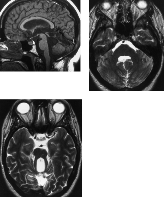
CASE 162 Upper Brainstem and Thalamic Cavernous Malformation

Comment

This case shows the typical appearance of an uncomplicated cavernous malformation involving the upper brainstem or cerebral peduncle and left thalamus. In the absence of edema, hemorrhage within the cavernous malformation is unlikely. The differentiation of a cavernous malformation from a hemorrhagic neoplasm on MR imaging occasionally can be difficult in the face of acute hemorrhage, particularly in the presence of edema and mass effect. Several imaging features may help to distinguish these two lesions. Findings favoring a cavernous malformation include focal heterogeneous high signal intensity, representing methemoglobin; a complete hypointense peripheral ring, representing hemosiderin, as in this case; and the absence of enhancing solid tissue. When all else fails, follow-up imaging in 4 to 6 weeks may be performed to assess for the expected temporal evolution of hemorrhage when secondary to a cavernous malformation.

Cavernous malformations may be present in up to 5% of the population. Most are located superficially in the cerebrum and are often closely associated with the adjacent subarachnoid space. They may occur deep within the cerebral hemispheres, although this is less common. Cavernous malformations occur less frequently in the infratentorial compartment. The most common brainstem location is the pons. Many are incidental asymptomatic lesions detected on imaging performed for other indications, as in this patient with traumatic injury. When lesions are symptomatic, symptoms may be related to lesion location or acute hemorrhage. In the cerebrum, the most common presentation is seizures. In the infratentorial compartment, neurologic deficits may occur on the basis of acute hemorrhage, thrombosis, or progressive enlargement of a cavernous malformation related to recurrent hemorrhages.

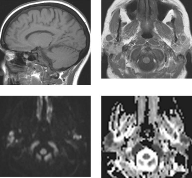
CASE 163 Capillary Telangiectasia and Developmental Venous Anomaly of the Brainstem

Comment

Capillary telangiectasias represent a cluster of abnormally dilated capillaries with intervening normal brain tissue. They usually represent clinically silent lesions that are detected on imaging studies acquired for unrelated reasons. Angiographically, they are most often occult. This case nicely illustrates the typical appearance of a capillary telangiectasia on MR imaging: a poorly demarcated region of “feathery” contrast enhancement, corresponding T2W images that show no significant associated signal abnormality, and gradient echo susceptibility images that show hypointensity in the lesion. It has been postulated that T2W shortening on gradient echo imaging may be related to intravascular deoxyhemoglobin from stagnant blood flow. In this case, regional cerebral blood volume shows increased flow that is believed to reflect pooling of blood in dilated capillaries and venules. Capillary telangiectasias may coexist with other vascular malformations, including cavernomas and DVAs.

DVAs are typically incidental vascular malformations representing an aberration in venous drainage. Within the venous network is intervening normal brain tissue, and no arterial elements are associated with these lesions. The angiomas are composed of a tuft of enlarged venous channels that drain into a common venous trunk, which then subsequently drains into the deep or superficial venous system. Typically, the lesions are clinically silent, although they may be associated with intracranial hemorrhage. These lesions have a characteristic MR imaging appearance, representing a cluster of veins oriented in a “radial” pattern that drain into a large central vein. There is usually no significant signal abnormality in the adjacent brain parenchyma. Angiographically, the arterial and capillary phases are normal, and there may be opacification of the DVA during the venous phase.

CASE 164 Central Neurocytoma

Comment

Central neurocytomas typically have a homogeneous cell population with neuronal differentiation. These benign neuroepithelial neoplasms occur in young and middle-aged adults. Patients may be asymptomatic, or may present with headache and signs of increased intracranial pressure, frequently due to hydrocephalus, as in this case. Central neurocytomas arise most commonly within the body of the lateral ventricle (less frequently, the third ventricle), adjacent to the septum pellucidum and foramen of Monroe. They have a characteristic attachment to the superolateral ventricular wall. Most are confined to the ventricles, although occasionally parenchymal extension may occur, as in this case, where there is growth into the frontal lobe. These features may help to distinguish neurocytomas from other intraventricular tumors, such as astrocytoma, giant cell astrocytoma, ependymoma, intraventricular oligodendroglioma, and meningioma. Preoperative diagnosis of central neurocytoma may help in planning therapy, because this tumor has a better prognosis than other intraventricular tumors arising in this area.

On CT and MR imaging, neurocytomas typically are heterogeneous masses that contain multiple cysts. They are well demarcated, with smooth, lobulated margins and moderate vascularity. Most neurocytomas have calcifications. On MR imaging, the more solid component of these tumors tends to follow the signal characteristics of gray matter. Signal voids may be related to calcification or tumor vascularity. Contrast enhancement is variable, ranging from none to moderate. On imaging and conventional pathologic evaluation (light microscopy), these tumors are frequently indistinguishable from oligodendrogliomas. The distinction between these two neoplasms is important because central neurocytomas have a more benign course and treatment may differ. Although neurocytomas have a favorable prognosis, malignant variants and recurrences may rarely occur.

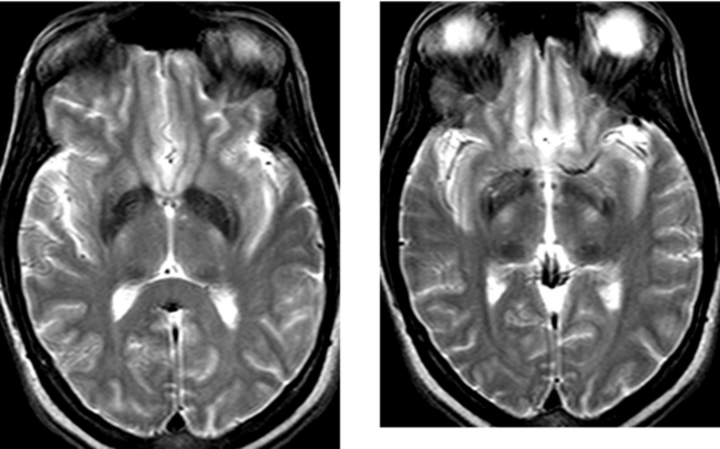
CASE 165 Marchiafava-Bignami Disease

Comment

Marchiafava-Bignami disease is a toxic demyelinating disorder initially described by two Italian pathologists. They identified it at autopsy in three patients with chronic alcoholism who presented in status epilepticus that subsequently progressed to coma. All three patients consumed large quantities of red wine. This has also been described in patients with significant nutritional deficiencies, and it has been described in other populations and with other alcoholic beverages. This diagnosis should be considered in patients with acute encephalopathy or progressive dementia and alcoholism. The disease may present acutely, with rapid deterioration, or may exist in a chronic form over a period of years. It is most commonly seen in men, and usually occurs in the third to fifth decade.

On pathologic evaluation, Marchiafava-Bignami disease is typified by demyelination and necrosis, and it occurs most commonly in the corpus callosum; however, there may be extensive demyelination involving multiple areas of the brain, as in this case, including the deep and periventricular white matter, as well as other commissural fibers. Occasionally, this may involve the subcortical white matter, but typically, the subcortical U-fibers are spared. Sagittal T1W images are valuable in assessing these patients because these images show the extensive callosal atrophy and the associated focal necrosis as demarcated regions of signal abnormality that are hypointense on T1W imaging and hyperintense on T2W imaging. This case nicely demonstrates all of these findings. The acute form of this disease presents with seizures, muscular hypertonia, dysphagia, and coma, and it is often fatal. In the acute form, diffusion-weighted images show hyperintensity (restricted diffusion), and there may be enhancement in affected regions of white matter. In the absence of a patient history, it would be difficult to distinguish this diagnosis from the other demyelinating processes that are more common, such as multiple sclerosis. However, Marchiafava-Bignami disease should be considered in patients with encephalopathy and a history of chronic alcoholism.

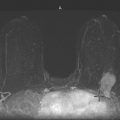
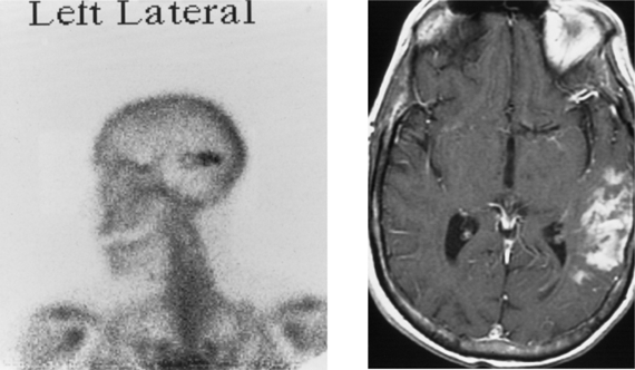
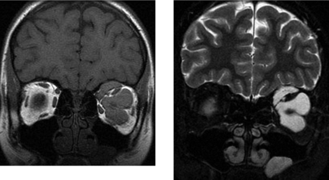
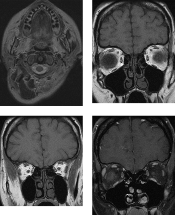
CASE 166 Metastatic Carcinoid to the Orbital Extraocular Muscles
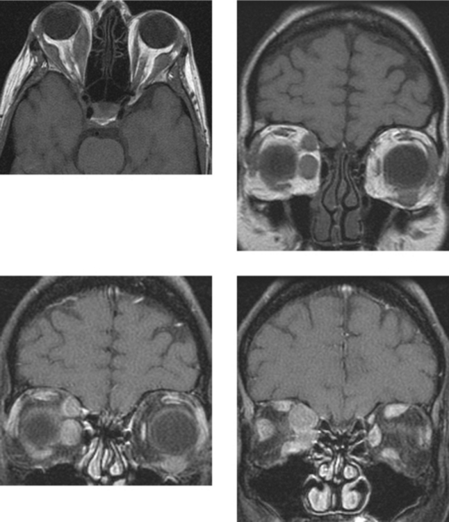
Comment

Carcinoid tumors arise from Kulchitsky cells that originate in the neural crest. Most carcinoids arise in the gastrointestinal tract or the lung. This case represents metastatic carcinoid to the orbit, specifically, to the extraocular muscles. With rare exception, the reported metastatic carcinoid tumors to the uvea all developed from primary bronchial carcinoids. In contrast, the vast majority of reported orbital metastases arose from ileal carcinoids. The metastatic potential of carcinoid tumors is related to the site of origin and to tumor size, with histologic features playing a lesser role. In symptomatic patients with intestinal carcinoid, more than 90% have metastatic disease, which is most common to the lymph nodes and liver. Tumors larger than 1 cm are more likely to metastasize.

Metastatic disease to the orbits is common. The imaging diagnosis of orbital metastases as the first manifestation of metastatic disease is not uncommon. For every case of clinically evident orbital metastases, there are several asymptomatic cases that go unrecognized. The most common location for orbital metastases is the globe, usually involving the region of the choroid and retina. Ocular metastases characteristically involve the uveoscleral region. The most common tumors to metastasize to the globe are breast and lung carcinoma in adults. Outside of the globe, orbital metastases most often are extraconal and are often related to bone metastases. Intraconal metastatic disease is usually related to direct extension of an ocular metastasis. Clinical manifestations of ocular metastases are variable. Some patients may be asymptomatic, whereas others may have proptosis, blurred vision, pain, or ophthalmoplegia, depending on the site of involvement.

CASE 167 Joubert’s Syndrome

Comment

Cerebellar developmental anomalies have been loosely categorized into complete or incomplete cerebellar agenesis, median aplasia or hypoplasia, and lateral aplasia or hypoplasia. These aberrations in cerebellar development may result in prominent CSF spaces or CSF collections or cystic dilation of the fourth ventricle (giant cisterna magna, Dandy-Walker malformations) in the posterior fossa. Other rarer entities are associated with enlargement or an abnormal configuration of the fourth ventricle. Such entities include aplasia or a spectrum of hypoplasias involving the cerebellar hemispheres, vermis, or brainstem. Unlike Dandy-Walker malformations, these cerebellar hypoplasias are not associated with posterior fossa cysts and less commonly have associated hydrocephalus or associated supratentorial anomalies.

The predominant abnormality in Joubert’s syndrome is aplasia or hypoplasia of the vermis, particularly the superior portion. In addition, these patients have dysplastic cerebellar tissue, including heterotopic and dysplastic cerebellar nuclei; abnormal development of the inferior olivary nuclei; and incomplete formation of the pyramidal decussation. It is an autosomal recessive disorder.

Magnetic resonance images in these patients show a characteristic appearance. Specifically, sagittal T1W images demonstrate a diminutive vermis. Axial images in particular show an enlarged fourth ventricle with a “bat-wing” shape. The superior cerebellar peduncles are vertically oriented and elongated in the anteroposterior direction. Because of the dysgenesis of the vermis, the hallmark of Joubert’s syndrome is separation or disconnection of the cerebellar hemispheres, which are apposed but not fused in the midline.

CASE 168 Systemic Non-Hodgkin’s Lymphoma

Comment

This case illustrates diffuse calvarial and skull base marrow replacement, in addition to solid bilateral parotid masses consistent with lymph nodes. The best diagnosis, based on all of the imaging findings, is non-Hodgkin’s lymphoma. In this case, lymph node and bone marrow biopsy both showed small B-cell lymphoma. Did you notice that the bilateral parotid masses show restricted diffusion (very suggestive of lymphoma)?

Magnetic resonance imaging is well suited to the evaluation of bone marrow because it can differentiate fat from other tissues. Signal intensity is directly related to relative amounts of fat, water, and cells in the marrow. Hematopoietic (red) marrow is composed of approximately 40% fat, 40% water, and 20% protein, compared with inactive fatty (yellow) marrow, which contains approximately 80% fat, 10% to 15% water, and 5% protein. On unenhanced T1W images, yellow marrow is hyperintense relative to muscle; it approaches the intensity of subcutaneous fat. Cellular red marrow has intermediate signal intensity, which may be isointense or slightly hyperintense relative to muscle, depending on the cell:fat ratio. Marrow conversion is a normal process in which yellow marrow gradually replaces red marrow. At birth, marrow is predominantly red in both the appendicular and axial skeletons. In the appendicular skeleton, most of the marrow has undergone conversion by the time an individual is 25 years of age. Residual red marrow is found in the proximal metaphyses of the femurs and humeri. In the axial skeleton, a larger portion of the marrow in adults remains hematopoietic compared with the appendicular skeleton.

A spectrum of processes may affect bone marrow. When demand for hematopoeisis is increased in response to systemic stresses, such as chronic anemia, heart failure, or heavy smoking, reconversion of fatty marrow to cellular marrow may occur in an anatomic pattern opposite to that of conversion. In addition, cellular replacement of bone marrow may occur in hematologic malignancies, metastatic disease, other infiltrative processes (such as granulomatous disease), and polycythemia vera.

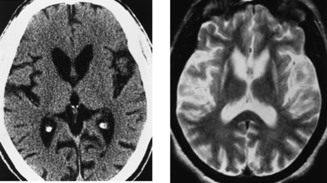
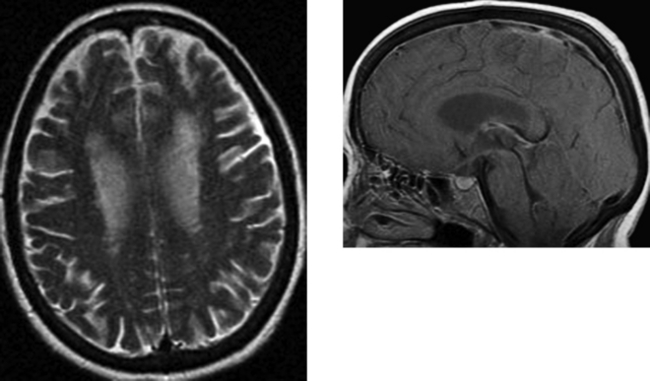
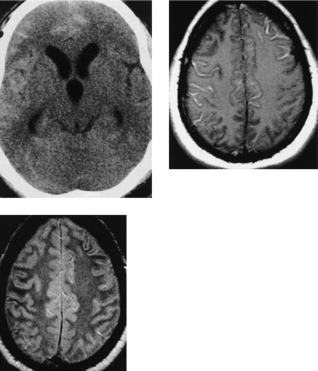
CASE 169 Abnormal Iron Deposition in Deep Gray Matter Nuclei

Comment

Detection of iron deposition within the deep gray matter of the brain (globus pallidus, putamen, caudate nucleus, thalamus), as well as the dentate nuclei, substantia nigra, and red nuclei, manifests as hypointensity on T2W images and may serve as an indirect marker of cerebral neurodegenerative processes. Axial T2W gradient echo susceptibility imaging shows striking hypointensity in involved structures due to paramagnetic effects. Although there is a spectrum of normal iron deposition within the basal ganglia and brainstem nuclei noted on pathologic examination and to some extent confirmed on MR imaging, certain trends are clear. Specifically, in healthy young adults, hypointensity within the substantia nigra, red nucleus, and dentate nucleus is seen on T2W images. Both evaluation of pathologic specimens and MR imaging suggest that the volume of iron products in these structures does not significantly increase with age. Although hypointensity in the globus pallidus is often present in young adults, the amount of hypointensity (reflective of iron deposition) increases with age. In addition, whereas hypointensity may be seen in the putamen, in normal subjects, this typically is not seen before the sixth decade. Of note, hypointensity in the thalamus or caudate nucleus generally is not present in normal subjects, regardless of age, and hypointensity in these locations is indicative of CNS disease.

A variety of CNS disorders may be suggested by the detection of hypointensity within the deep gray matter and nuclei that is different from the expected normal patterns. Increased brain iron levels may be seen in a variety of pathologic states, including demyelinating or dysmyelinating diseases (multiple sclerosis), the leukodystrophies, brain anoxia and infarction, movement disorders (Wilson’s disease, Hallervorden-Spatz disease), endocrinologic or metabolic disorders (hyperparathyroidism, hypoparathyroidism, pseudohypoparathyroidism), and significant closed head trauma. The patient in this case has underlying amyotrophic lateral sclerosis (Lou Gehrig’s disease).

CASE 170 Herpes Encephalopathy—Human Herpesvirus-6 Infection after Organ Transplantation

Comment

Human herpesvirus-6 (HHV-6) is a double-stranded DNA virus. More than 90% of the general population is seropositive for HHV-6. It is excreted by the salivary glands and may be passed to infants from their mothers. HHV-6 has a strong affinity for the CNS, and has been detected by polymerase chain reaction in up to one third of normal brain specimens, suggesting that the brain might be a latent viral site. Recently, HHV-6 encephalopathy has been reported in immunocompromised patients, especially patients who have undergone hematopoietic stem cell or solid-organ transplantation (lung and liver). Infection has typically been identified within 4 weeks of the transplantation. The pathogenesis is considered to be reactivation of the recipient’s latent HHV-6 infection, and not infection from the donor. Immunocompromised patients are at risk for a spectrum of disease processes that may affect the CNS, and their symptoms are frequently nonspecific. Common neurologic symptoms in HHV-6 infection include disorientation, confusion, and short-term memory loss. Coma, hypopnea, and seizures have been reported.

Early MR imaging findings, as in this case, include high signal intensity on FLAIR, T2W and diffusion-weighted images of the mesial temporal lobe structures (hippocampus and amygdala). The diffusion-weighted abnormality is accompanied by hypointensity on apparent diffusion coefficient maps. Enhancement usually is not present. In transplantation, acyclovir is routinely administered to prevent reactivation of herpes viruses. However, acyclovir is not effective against HHV-6 because it lacks virus-specific thymidine kinase. Gancyclovir and foscarnet can be effective against HHV-6, but serious side effects, including myelosuppression and nephrotoxicity, may occur. Therefore, these drugs are not usually given prophylactically. Early diagnosis is critical to prevent serious neurologic sequelae. Mesial temporal involvement seen on MR images in a transplant recipient receiving preventive treatment with acyclovir is highly suggestive of HHV-6–associated encephalopathy.

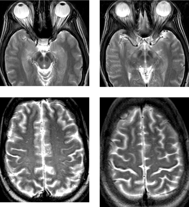
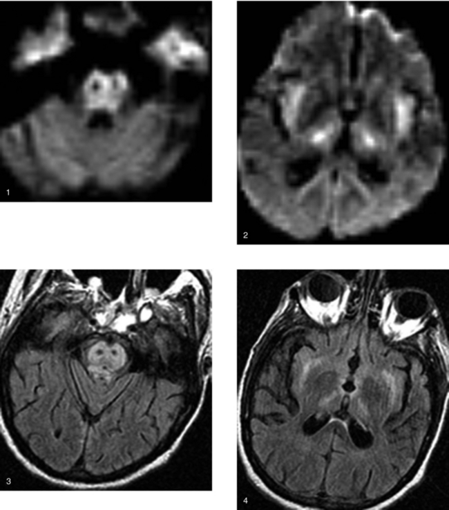
CASE 171 Jakob-Creutzfeldt Disease

Comment

Jakob-Creutzfeldt disease is rare and is caused by a prion agent composed of protease-resistant protein that affects the CNS and results in rapid, progressive neurodegeneration. Approximately 1 in every 1 million persons worldwide is infected. It is thought to be caused by a slow virus, an organism devoid of active nucleic acid. The most common clinical presentation is that of rapidly progressive dementia. Other neurologic symptoms include upper motor neuron signs, ataxia, myoclonus, and sensory deficits. The characteristic diagnostic triad of progressive dementia, myoclonic jerks, and periodic sharp-wave electroencephalographic activity is present in approximately 75% of cases. Prognosis is poor, with death usually occurring within 1 year from the onset of symptoms. Histologic evaluation shows neuronal degeneration and gliosis in the gray matter, especially the cortex, but also in the deep gray matter of the corpus striatum and thalami, as in this case. Spongiform changes are characteristic. Inflammatory changes are usually not present. The disease is best known in association with mad cow disease (bovine spongiform encephalopathy) in the United Kingdom; however, there have been scattered sporadic cases described with corneal transplantation as well as with implantation of cerebral electrodes.

Computed tomography may show no abnormality; however, atrophy (most commonly cortical) is the next most common presentation. On MR imaging, in addition to cortical atrophy, T2W hyperintensity in the deep gray matter, especially the caudate and putamen nuclei, but also the thalami, is observed. Lesions typically are bilateral and are not associated with enhancement or significant mass effect. In addition, abnormalities involving the gray and white matter have been noted within the cerebral hemispheres. Several reported cases of sporadic Creutzfeldt-Jakob disease have shown increased signal intensity in the basal ganglia or cerebral cortex on diffusion-weighted images (reduced diffusion), as in this case. Ribbon-like areas of hyperintensity in the cerebral cortex on diffusion-weighted images have also corresponded to the localization of periodic sharp-wave complexes on electroencephalogram.

CASE 172 Orbital Lymphangioma

Comment

The most common vascular neoplasms in the orbit include cavernous hemangioma, capillary hemangioma, and lymphangioma. They are usually distinguishable based on their location in the orbit, their imaging appearance, and the age of the patient.

Capillary hemangiomas are seen almost exclusively in children and usually present clinically within the first months of life. They usually peak in size within the first 18 months of life and then often undergo spontaneous regression. They are infiltrative lesions that may extend into the periorbita, eyelid, and conjunctiva. Because of their vascularity, they may increase in size during crying or coughing (Valsalva maneuvers). Capillary hemangiomas may involve any portion of the orbit and are frequently associated with vascular flow voids on MR imaging. Cavernous hemangiomas are the most common retrobulbar mass in adults. Their imaging appearance is distinctly different from that of capillary hemangiomas. These are demarcated lesions that appear hyperdense on unenhanced CT, hypointense on T1W imaging, and markedly hyperintense on T2W imaging. After contrast, they enhance avidly. Although phleboliths may be present, this finding is not as common as with hemangiomas in other parts of the body.

Lymphangiomas are poorly demarcated, infiltrating lesions that are less common than hemangiomas. They are usually extraconal in origin and may extend to involve the adjacent extraocular muscles, the soft tissues in the periorbital region, and the conjunctiva. Less commonly, they may extend to the intraconal compartment, as in this case. Small lesions may be asymptomatic; however, patients may become acutely symptomatic as a result of hemorrhage within the lesion. On MR imaging, lymphangiomas have a characteristic appearance. They are usually multilobular, with regions of cystic change and peripheral enhancement. Hemorrhage within cystic regions of the neoplasm is common and usually appears hyperintense on T1W imaging. When present, a key to the diagnosis is the finding of fluid–hemorrhage levels within the lesion. Because lymphangiomas are not encapsulated, surgical resection is difficult and recurrence common.

CASE 173 Carotid–Cavernous Fistula—Part I

Comment

There are two basic types of carotid–cavernous vascular malformations, direct (type A) and indirect (dural, types B–D), each of which has a different etiology. Carotid–cavernous fistulas represent a direct communication between the intracavernous portion of the internal carotid artery and the cavernous venous sinus. The typical clinical presentation of a carotid–cavernous fistula is ophthalmologic symptoms, including pulsatile proptosis, pain, chemosis, and orbital bruit. This is because the cavernous sinus directly communicates with the ophthalmic veins, and an abnormal shunt between the sinus and the internal carotid artery can transmit arterial pressure to these veins. In addition, arterial perfusion to the globe is decreased, leading to visual loss. Direct carotid–cavernous fistulas are most commonly the result of head trauma; however, spontaneous carotid–cavernous fistulas may be seen in a spectrum of disorders, including atherosclerosis in the elderly, rupture of a carotid–cavernous aneurysm, or in association with underlying vascular dysplasias. An indirect carotid–cavernous vascular malformation, otherwise known as a dural arteriovenous fistula, is a shunt between meningeal branches of the cavernous internal carotid artery (type B), meningeal branches of the external carotid artery (type C), or meningeal branches of both the intracavernous carotid artery and the external carotid artery (type D) with the cavernous venous sinus.

The clinical presentation and imaging findings are normally diagnostic of a direct carotid–cavernous fistula. CT and MR imaging often show enlargement of the superior ophthalmic vein, cavernous sinus, or petrosal venous plexus, as in this case. Proptosis, periorbital soft tissue swelling, and diffuse enlargement of the extraocular muscles are commonly present. MR angiography and catheter angiography show direct communication between the cavernous internal carotid artery and the cavernous venous sinus as well as early filling of the ipsilateral cavernous sinus, superior or inferior ophthalmic veins, and petrosal venous complex. In high-flow lesions, the contralateral venous system may opacify, as in this case, through both intercavernous veins and the petrosal venous complex.

CASE 174 Carotid–Cavernous Fistula—Part II

Comment

In this case, the left carotid injection shows the site of the fistula between the cavernous left internal carotid artery and the cavernous venous sinus. Contralateral injection of the right carotid artery and injection of the vertebral basilar arterial system are important in evaluating for collateral flow, particularly if the involved internal carotid artery must be sacrificed to close the fistula, as was necessary in this case. Right common carotid artery injection in this patient shows a patent anterior communicating artery with cross-filling of the left anterior and middle cerebral circulation. Left vertebral artery injection shows filling of the left supraclinoid internal carotid artery through a patent posterior communicating artery.

Management of these lesions in the majority of cases is usually with interventional neuroradiologic procedures. Symptomatic direct carotid–cavernous fistulas (type A) spontaneously resolve only in rare cases. The goal of treatment is to eliminate flow through the fistula as well as to maintain internal carotid patency when possible. Treatment of direct and indirect fistulas may differ. Direct carotid–cavernous fistulas are usually treated transarterially with detachable coils or balloon embolization, with flow directed through the fistula into the cavernous sinus, tamponading the hole in the internal carotid artery. In the event that a transarterial route is not possible or is ineffective, a transvenous approach using platinum coils may be warranted. This can be achieved either via the femoral route or surgically via the superior ophthalmic vein. Complicated carotid–cavernous fistulas, residual carotid–cavernous fistulas after embolization with an arterial approach, and dural arteriovenous fistulas of the cavernous sinus may sometimes need to be treated transvenously. Gamma knife radiosurgery has also been shown to be effective in treating indirect dural arteriovenous fistulas.

CASE 175 Gliomatosis Cerebri

Comment

This case illustrates the typical radiologic appearance of gliomatosis cerebri. There is extensive abnormality within the brain that affects the white matter; however, the gray matter is also involved. This extensively infiltrative process affects large portions of the cerebrum, including the right temporal, occipital, and frontal lobes. There is mild gyral swelling and sulcal effacement. There is extension across the splenium of the corpus callosum that is mildly expanded, as well as the anterior commissure, with involvement of the left cerebrum to a lesser extent. There are no regions of necrosis, there is no circumscribed mass, and there is no enhancement (not shown). Also, on image 1, note the involvement of the optic chiasm and bilateral optic tracts.

Gliomatosis cerebri is characterized by an extensive infiltrative pattern throughout the involved portions of the brain disproportionate to the remainder of the histologic findings, including a relative paucity of cellularity, anaplasia, and necrosis. In addition to the disproportionate histologic findings relative to the degree of infiltration seen on MR imaging, clinical symptoms are characteristically mild relative to the degree of brain involvement. Patients often present only with altered mental status or a change in personality. Headaches and seizures may occur. Focal neurologic deficits occur late in the course of disease. Although this tumor may affect patients of any age, it most commonly presents between the third and fifth decades. In addition to the extensive parenchymal involvement and the signal alteration seen on MR imaging, other findings that may suggest the diagnosis include mild diffuse sulcal and ventricular effacement. Not uncommonly, a large resection or lobectomy is necessary to make the diagnosis pathologically because biopsy may provide insufficient material. Therefore, recognition of the imaging findings, in combination with the patient’s history, is important in diagnosing this neoplasm. Radiologists play a critical role by suggesting the diagnosis, mapping the extent of disease, and guiding biopsy.

CASE 176 Canavan Disease

Comment

The leukodystrophies, or dysmyelinating disorders, represent a spectrum of inherited diseases that usually result in both abnormal formation and abnormal maintenance of myelin. Many of the more common of these rare disorders are inherited in an autosomal recessive pattern. In many of the leukodystrophies, specific enzyme deficiencies have been identified as the cause. These diseases cause abnormal growth or development of the myelin sheath, the fatty covering that acts as an insulator around nerve fibers in the brain. Myelin is made up of at least 10 different chemicals. Each of the leukodystrophies affects one (and only one) of these substances.

Canavan disease is transmitted as an autosomal recessive disorder usually identified in infants of Ashkenazi Jewish descent and in Saudi Arabians. It is the result of a deficiency of N-acetylaspartoacyclase. Infants may have macrocephaly due to enlargement of the brain. On histologic evaluation, there is diffuse demyelination and the white matter is replaced by microscopic cystic spaces, giving it a “spongy” appearance. In contrast to most of the other dysmyelinating syndromes, Canavan disease preferentially begins in the subcortical white matter and later spreads to diffusely involve the deep white matter. There may be sparing of the internal capsules. There may be bilaterally symmetric T2W signal abnormality in the deep gray matter, as in this case. The brainstem is involved in late disease. Usually, the ventricles remain normal or may be slightly small; however, in the late stages of disease, when cerebral atrophy occurs, there may be proportionate enlargement of the ventricles and cerebral sulci. Diffusion-weighted imaging shows diffuse restriction, and spectroscopy shows high levels of N-acetylaspartoacyclase.

Alexander disease is different from many of the leukodystrophies in that no familial pattern has been recognized. Like Canavan disease, it presents with macrocephaly in addition to developmental delay and spasticity. The deep white matter is usually involved early, and the internal capsules are typically involved (in contrast to Canavan disease, in which they are often relatively spared).

CASE 177 Neurofibromatosis Type 2

Comment

This case shows imaging findings characteristic of neurofibromatosis type 2, including bilateral vestibular schwannomas, as well as multiple dural-based, extra-axial, avidly enhancing masses, consistent with meningiomas.

Neurofibromatosis type 2 is an autosomal dominant disorder transmitted on chromosome 22. It typically presents in adolescence or young adulthood, and the most common clinical presentation is bilateral sensorineural hearing loss as a result of vestibular schwannomas. The radiologic hallmark of neurofibromatosis type 2 is the presence of bilateral vestibular schwannomas. Otherwise, the diagnosis of neurofibromatosis type 2 can be made if a patient has a unilateral vestibular schwannoma and a first-degree relative with neurofibromatosis type 2, or if patient has a first-degree relative with neurofibromatosis type 2 and at least two schwannomas, meningiomas, or ependymomas. Unlike neurofibromatosis type 1, cutaneous manifestations in neurofibromatosis type 2 are rare. CNS tumors are present in virtually 100% of patients with neurofibromatosis type 2. Schwannomas most commonly involve cranial nerve VIII. The trigeminal nerve is the next most common site for schwannomas. Although most of these tumors are sporadic, those arising from more than one cranial nerve or from cranial nerves III through VI should prompt a search for neurofibromatosis type 2. Other imaging findings that may be present in neurofibromatosis type 2 include prominent calcifications along the choroid plexus, or occasionally along the cerebral or cerebellar cortex. Lesions within the spinal canal are common and include schwannomas and meningiomas. Intramedullary tumors are typically ependymomas.

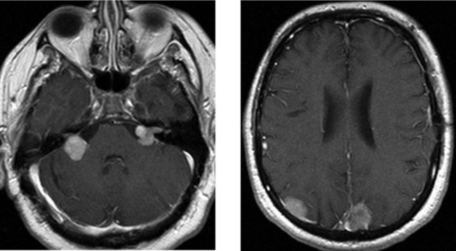
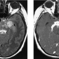
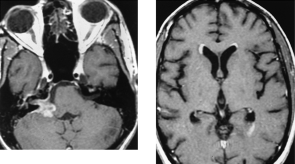
CASE 178 Metastasis to the Cerebellopontine Angle—Ocular Melanoma

Comment

This case shows a poorly demarcating enhancing mass in the right cerebellopontine angle, with extension into the internal auditory canal (IAC). The differential diagnosis includes schwannoma, meningioma, metastatic disease, and an inflammatory mass. The appearance of the mass is atypical for a meningioma, given the extension into the IAC and the poorly demarcated margins. Similarly, poorly defined boundaries, lack of cystic degeneration, and absence of expansion of the IAC, in conjunction with abnormality within it, are atypical features of vestibular schwannoma.

Malignant melanoma of the uveal tract represents the most common intraocular malignancy of adults. It is usually unilateral and commonly presents in the fifth and sixth decades. On CT, ocular melanomas are typically hyperdense relative to the vitreous and enhance after contrast. On MR imaging, melanotic melanomas are characteristically hyperintense on T1W and hypointense on T2W images. The signal characteristics are related to the paramagnetic effects of melanin. In the absence of intravenous contrast, melanoma could be mistaken for hemorrhage, such as that seen with retinal detachment. However, enhanced images typically show solid enhancing tissue with melanomas. Did you notice the primary melanoma of the right uveal tract?

The most common site of metastases to the orbit is also along the uveal tract, and for this reason, metastatic disease to the orbit may be confused with primary ocular melanoma. Metastatic mucin-secreting adenocarcinomas of the uveal tract have been noted to have signal characteristics similar to those of ocular melanoma.

The most important role of MR imaging is in detecting extraocular extension of primary ocular melanoma. Episcleral involvement, retrobulbar extension, and occasional perineural and intracranial subarachnoid spread may occur. Systemic metastases are most commonly noted in the liver and lungs, but may also be seen in the bone and brain. Therefore, a careful metastatic workup is required to prevent unnecessary enucleation. Two treatments for primary ocular melanoma include removal of the eye (enucleation) and irradiation. Recent studies suggest similar survival rates for these treatments.

CASE 179 Retained Gadolinium—Renal Failure

Comment

Fluid-attenuated inversion recovery is routinely used in brain imaging due to its high lesion-to-tissue contrast from T2 prolongation and nulling of the normal CSF background. Therefore, any alteration in the CSF results in increased signal intensity in the CSF. Flair imaging is particularly sensitive for pathologic entities that affect the CSF in the subarachnoid spaces. Hyperintense CSF in the subarachnoid spaces on FLAIR imaging has been reported in numerous conditions, including subarachnoid hemorrhage, meningitis, meningeal carcinomatosis, venous sinus thrombosis, status epilepticus, and stroke. Increased signal intensity in the subarachnoid spaces can be a diagnostic pitfall in patients receiving supplemental oxygen during MR imaging of the brain and in patients with renal insufficiency who have previously received gadolinium (as in this case).

Gadolinium has also been reported to diffuse across the choroid plexus and uveochoroid membrane in patients with impaired renal function, resulting in FLAIR hyperintensity in the ventricular system and in the vitreous and aqueous humor of the eye. The mechanism by which CSF diffuses into the CSF spaces is not clearly understood. It has been postulated that in patients with renal insufficiency, the gadolinium may move across an osmotic gradient at the circumventricular organs in the setting of prolonged elevation of plasma concentrations because it is primarily cleared through glomerular filtration. Some patients will have undergone gadolinium-enhanced MR imaging 24 to 48 hours before MR imaging of the brain. The radiologist should be aware that delayed gadolinium chelate clearance can cause increased signal intensity in the CSF on FLAIR imaging in patients with or without renal failure and without abnormalities known to disrupt the blood–brain barrier.

CASE 180 Amyotrophic Lateral Sclerosis—Lou Gehrig’s Disease

Comment

Amyotrophic lateral sclerosis (Lou Gehrig’s disease) is the most common of the neurodegenerative disorders involving the motor neurons. It occurs in approximately 1 in every 100,000 people annually. Most cases are sporadic, although autosomal dominant transmission may occur. Amyotrophic lateral sclerosis typically presents in the sixth decade of life; clinical manifestations include hyperreflexia, weakness of the hands and forearms, spasticity, and cranial neuropathies. The hypoglossal nerve is most commonly affected, and its involvement may be detected on imaging as denervation atrophy with fatty replacement of the tongue.

Amyotrophic lateral sclerosis typically involves the corticospinal tracts and motor neurons. Progression is usually relentless, with death frequently occurring within 3 to 6 years of disease onset. The cause is unknown. In extreme cases, abnormal T2W signal intensity may extend from the cortex, along the precentral gyrus of the motor strip (pyramidal Betz’s cells or upper motor neurons); through the corona radiata, the posterior part of the posterior limb of the internal capsule, the cerebral peduncles, and brainstem; and down to the ventral and lateral portions of the spinal cord. Abnormal T2W hypointensity, believed to be related to the deposition of iron or other minerals, may be present along the cerebral cortex in the motor strip, as in this case. SPECT with N -isopropyl-p-I123 iodoamphetamine may show decreased uptake in the cerebral cortex, including the motor cortex. Spectroscopy of the precentral gyrus region has shown a strong correlation between reduced N-acetylaspartate and glutamate levels and elevated choline and myoinositol levels and severity of disease. MR imaging of the spinal cord may show atrophy along the corticospinal tracts.

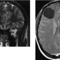
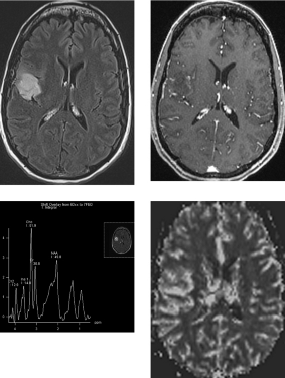
CASE 181 High-Grade Anaplastic Astrocytoma

Comment

This case shows abnormal signal intensity in the superficial cortex and subcortical white matter of the inferior right frontal lobe and superior right temporal lobe. There is mild local mass effect manifested by gyral expansion and sulcal effacement. No avid contrast enhancement is identified. These features on conventional FLAIR and gadolinium-enhanced images suggest a low-grade astrocytoma. On proton MR spectroscopy, an increased choline peak is highly suggestive of a malignant neoplasm. Importantly, perfusion imaging shows markedly elevated regional cerebral blood volume, also highly suggestive of a high-grade astrocytoma.

There are two classes of astrocytic tumors: those with narrow zones of infiltration (pilocytic astrocytoma, subependymal giant cell astrocytoma, pleomorphic xanthoastrocytoma) and those with diffuse zones of infiltration. According to the WHO classification, infiltrating astrocytic tumors may be divided into three subtypes: low-grade astrocytoma, anaplastic astrocytoma, and GBM. The histologic criteria for these subdivisions depend on the cellular density, number of mitoses, presence of necrosis, nuclear and cytoplasmic pleomorphism, and vascular endothelial proliferation. GBM typically has all of these histologic features, whereas low-grade astrocytomas may only demonstrate minimal increased cellularity and cellular pleomorphism. The presence of necrosis and vascular endothelial proliferation in particular favors GBM.

Histologically, anaplastic astrocytomas have features that are between those of a low-grade astrocytoma and those of GBM. On imaging, they typically show heterogeneous enhancement; however, the enhancement pattern may vary from avid and nodular to minimal (as in this case). Necrosis is much less common than that seen in the more malignant GBM. Anaplastic astrocytomas are highly malignant, with an average survival time of 2.5 years after diagnosis.

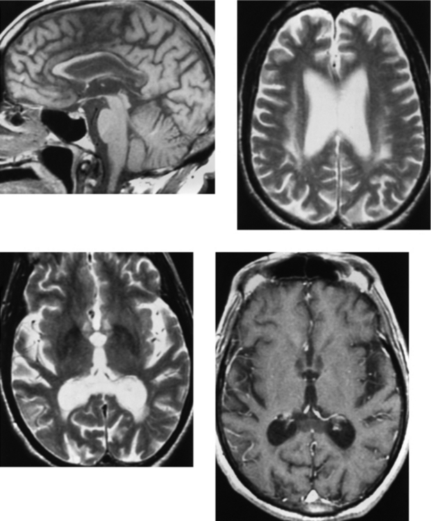
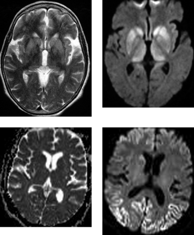
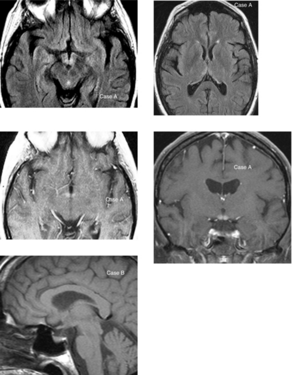
CASE 182 Wilson’s Disease

Comment

Wilson’s disease (hepatolenticular degeneration) is an autosomal recessive hereditary disease that results from abnormal metabolism of copper caused by deficiency of its carrier protein, ceruloplasmin. As a result, there is extensive abnormal deposition of copper in multiple organ systems (most pronounced in the liver and brain). Although neurologic symptoms may be directly related to copper deposition within the brain parenchyma, they may also be a manifestation of hepatic encephalopathy caused by liver failure. Neurologic signs and symptoms may include a pseudoparkinsonian-like syndrome, with rigidity, gait disturbance, and difficulty with fine motor skills. Dysarthria and progressive cognitive and psychiatric disturbances may also be present. Symptoms usually begin at 10 to 20 years of age, but sometimes do not begin until the age of 30 years and beyond. Presentation before 5 years of age is rare, even though the biochemical defect is present at birth. Treatment of Wilson’s disease is with D-penicillamine (chelation therapy).

On MR imaging, the most common finding may be atrophy. Regions of abnormal T2W signal intensity in particular are noted to involve the deep gray matter of the basal ganglia and the thalami, as well as the white matter. In particular, the putamen of the lentiform nucleus and the caudate are involved. In addition to signal abnormality, atrophy of the caudate nuclei may be present. Regions of T2W hypointensity have also been noted in the basal ganglia and have been attributed either to the paramagnetic effects of copper or possibly to associated iron deposition. Abnormalities on MR imaging may also be present in the brainstem and in the white matter of the cerebellum, as in this case. The diagnosis is made by laboratory analysis in which there are elevated copper levels within the urine as well as low serum ceruloplasmin levels.

CASE 183 Pleomorphic Xanthoastrocytoma

CASE 184 Primary Leptomeningeal Melanosis

Comment

Leptomeningeal melanosis refers to an increase in the number of melanocytes in the leptomeninges. Melanoblasts (precursors to melanocytes) are derived from the neural crest. Proliferation of melanocytes along the leptomeninges may be seen in a variety of forms, including primary leptomeningeal melanosis, primary leptomeningeal melanoma, and metastatic melanoma. Metastatic melanoma is the most common cause of such proliferation.

The diagnosis of primary leptomeningeal melanosis can be made when the following criteria are met: (1) there is a proliferation of melanocytes within the meninges or melanocytosis (high melanocyte count in the CSF); (2) there is no cutaneous or ocular melanoma; and (3) an extensive workup, including a bone scan as well as CT of the chest, abdomen, and pelvis, shows no metastatic lesions. Primary CNS melanocytic proliferation carries a poor prognosis. Patients may present with signs of increased intracranial pressure, seizures, and cranial neuropathies. Leptomeningeal melanosis may be benign or malignant. When malignant, there is characteristically invasion of the adjacent brain or the spinal cord.

Because melanocytosis in the CSF may be associated with a high protein content, unenhanced CT may show a hyperdense exudate in the subarachnoid spaces that may be mistaken for acute hemorrhage. On MR imaging, unenhanced T1W and FLAIR images may show hyperintense lesions along the leptomeninges within the subarachnoid spaces. Involved leptomeninges and brain may show regions of T2W hypointensity because of the paramagnetic effects of melanin. Involved leptomeninges show prominent enhancement.

Leptomeningeal melanosis associated with cutaneous nevi is referred to as neurocutaneous melanosis and is classified as a phakomatosis. These patients typically have multiple congenital hyperpigmented or giant hairy pigmented cutaneous nevi. Malignant transformation of meningeal melanosis may occur in as many as 50% of patients and often presents with seizures when the intracranial compartment is involved because of invasion. Leptomeningeal melanosis responds poorly to radiation and chemotherapy. Reported cases suggest that intrathecal recombinant interleukin-2 may provide a more promising response.

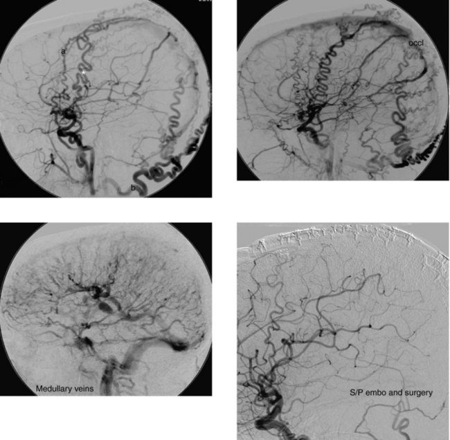
CASE 185 Dural Arteriovenous Fistula—Part I

Comment

Dural arteriovenous malformations (DAVMs) and fistulas account for approximately 12% of all intracranial vascular malformations. More than half occur in the posterior fossa, and they may be categorized based on the dural venous sinus involved by the vascular abnormality. These vascular malformations most commonly involve the sigmoid or transverse sinuses, accounting for as many as 70% of all of these lesions. The cavernous sinus is the next most common site for DAVMs and DAVFs, representing approximately 10% to 15% of these lesions.

Most dural vascular malformations present clinically in older adults. They are believed to be acquired lesions that typically arise as a consequence of dural venous sinus thrombosis. As a result, a collateral network of vessels develops, including enlargement of normally present microscopic arteriovenous shunts within the dura. In addition, as arteriovenous shunting increases, venous hypertension develops. In certain DAVMs, venous hypertension may result in retrograde filling of leptomeningeal or cortical veins that communicate with the involved sinus, as in this case. In such cases, rupture of the enlarged venous collaterals may occur. Approximately 10% to 15% of DAVMs are associated with intracranial hemorrhage, which is usually intraparenchymal or subarachnoid. Hemorrhage is associated with lesions in which there is development of dilated leptomeningeal veins. DAVMs and DAVFs that drain strictly to the dural venous sinuses are not usually associated with hemorrhage.

It is important to recognize that DAVMS and DAVFs are difficult to diagnose on CT and MR imaging. Imaging findings may include venous thrombosis, dilated cortical veins, or intracranial hemorrhage. However, imaging findings may be normal, and a high index of suspicion is required.

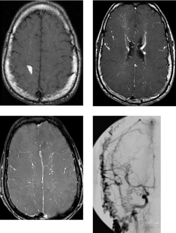
CASE 186 Dural Arteriovenous Fistula—Part II

Comment

The right common carotid injection shows that the external artery branches fill ahead of the right anterior and middle cerebral arteries, consistent with excessive flow to the external carotid circulation. There is a dural fistula with the superior sagittal sinus that is supplied by multiple feeders, including the middle meningeal, superficial temporal, occipital, and posterior auricular arteries. The superior sagittal sinus is interrupted or occluded at the posterior two thirds (occl ), with arteriovenous shunting into the anterior and posterior components of the superior sagittal sinus around the obstruction. There is retrograde filling into enlarged cortical and medullary veins in both cerebral hemispheres on the delayed venous phase. Delayed drainage does occur into an enlarged deep venous system that drains into the transverse and sigmoid sinuses. Left common carotid arteriography showed similar findings.

The superficial temporal, middle meningeal, and occipital artery feeders to the dural nidus were embolized using endovascular techniques, with significant occlusion of the nidus. Subsequently, surgery was performed. Angiography performed after embolization and surgery showed excellent treatment of the DAVM or fistula, with a normal-appearing angiogram.

This patient presented with progressive dementia, a less common presentation of DAVM. The dementia syndrome (venous hypertensive encephalopathy) is secondary to elevated intracranial venous pressure with associated diminished cerebral perfusion. This type of dementia is potentially reversible, as it was in this case, after endovascular embolization and neurosurgical resection. This patient experienced significant return of cognitive function.

CASE 187 Fetal Schizencephaly

Comment

Fetal MR imaging is an increasingly available technique used to evaluate the fetal neural axis. Ultrafast T2W sequences, parallel imaging, and new coil designs have allowed fetal MR imaging to be used to evaluate processes that cannot be evaluated by other imaging techniques. Fetal MR allows assessment of in vivo brain development and early diagnosis of congenital abnormalities inadequately evaluated by prenatal sonography. It has been especially useful in the evaluation of sonographically diagnosed ventriculomegaly, suspected abnormalities of the corpus callosum, and posterior fossa lesions.

Schizencephaly is a migrational abnormality that results from an injury to the germinal matrix and causes failure of normal migration and neuronal differentiation. This anomaly is thought to be the result of an in utero watershed ischemic event leading to damage not only to the germinal matrix but also to the radial glial fibers along which the neurons normally migrate from the germinal region to their final destination in the cortex. As a result, schizencephaly extends from the ventricular surface to the subarachnoid surface of the brain and is lined by dysplastic gray matter. Usually, there is a CSF cleft between the layers of gray matter. When the CSF cleft is large and gaping, it is referred to as “open-lip” schizencephaly. When the layers of gray matter are in apposition or when there is only a thin layer of CSF between them, the condition is referred to as “closed-lip” schizencephaly.

Up to 25% to 50% of patients with schizencephaly have septo-optic dysplasia. Patients with migrational anomalies usually present with a seizure disorder. Depending on the size of the migrational abnormality, there may also be focal neurologic deficits, such as hemiparesis. MR imaging is the imaging modality of choice to assess these abnormalities. Closed-lip schizencephaly, when the gray matter layers are apposed, may be difficult to detect. A small ventricular “dimple” or diverticulum may be a clue to the diagnosis and should prompt a close search for closed-lip schizencephaly. Other conditions that may cause dimpling of the ventricle are previous injuries, such as periventricular ischemia or infection.

CASE 188 Wernicke Encephalopathy

Comment

These cases show many of the radiologic findings seen in Wernicke encephalopathy. Case A shows FLAIR high signal intensity and enhancement in the mamillary bodies and FLAIR high signal intensity in the medial thalami. Wernicke encephalopathy is typically associated with generalized cerebral cortical and cerebellar vermial atrophy. In addition, specific deep structures are involved and are best assessed with MR imaging, which is more sensitive than CT in evaluating the small structures involved in this entity. Abnormal T2W and FLAIR hyperintensity is seen in the mamillary bodies (in essentially all patients), and may also be seen in the hypothalamus, periaqueductal gray matter, and medial thalami. Imaging findings are often bilaterally symmetric. In the acute setting, there may be mild swelling associated with the signal alteration, and enhancement has also been reported, as is seen in Case A. Resolution of the signal alterations after treatment with thiamine has been reported. In the late stages, atrophy (particularly of the mammillary bodies) may be the main finding, as is seen in Case B.

Wernicke encephalopathy is related to thiamine deficiency and is found most commonly in chronic alcoholism; however, this vitamin deficiency may also be present in other conditions that result in chronic malnutrition, such as anorexia nervosa, prolonged infectious or febrile conditions, and hyperemesis gravidarum. Wernicke syndrome has also been reported in association with long-term parenteral therapy. Wernicke encephalopathy clinically manifests with tremors, ocular symptoms (nystagmus and gaze paralysis), confusion, delusions, and ataxia; in contrast, Korsakoff psychosis is manifested by retrograde amnesia and difficulty acquiring new information. The two sometimes occur together.

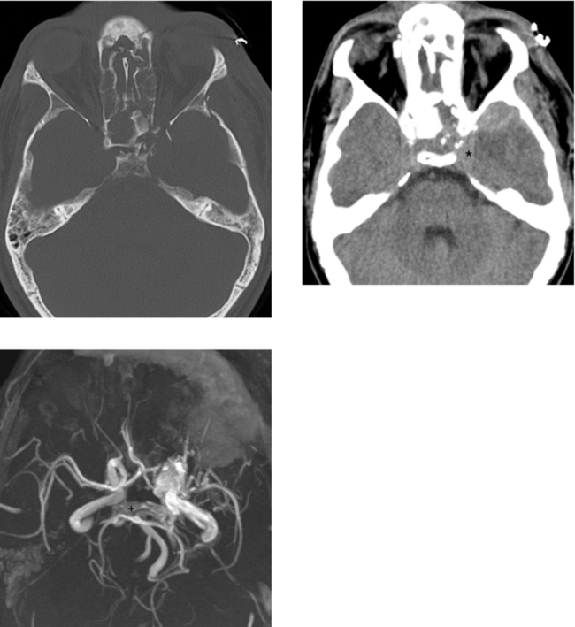
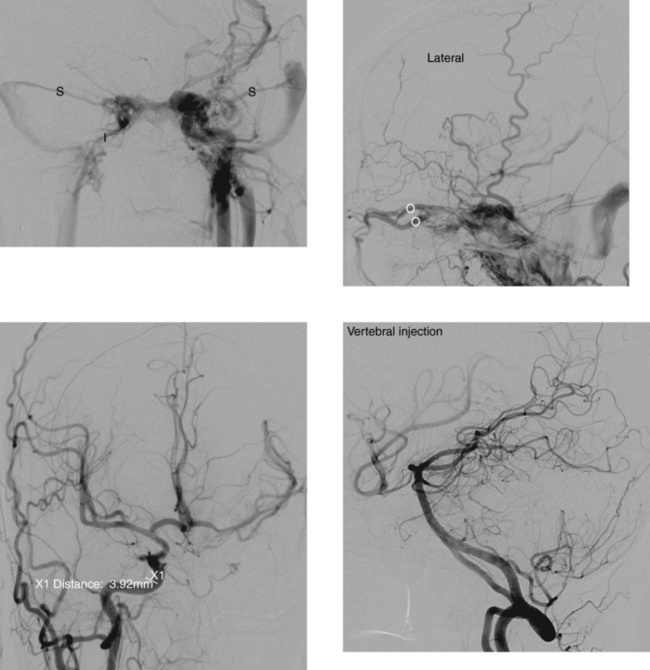
CASE 189 Ruptured Mycotic Aneurysm
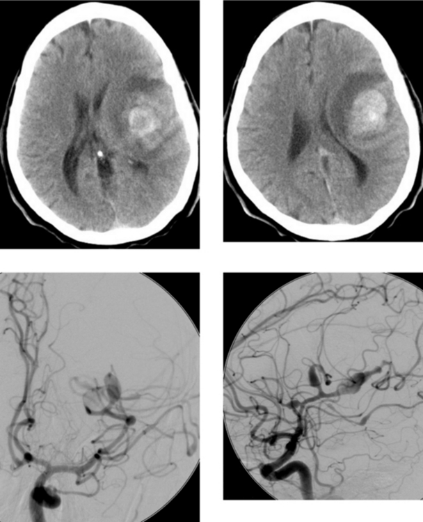
Comment

Up to 15% of patients with acute subarachnoid hemorrhage die before reaching the hospital. The incidence of aneurysmal rebleeding occurs at a rate of approximately 2% per day in the first 2 weeks after the initial hemorrhage. There are several issues that the angiographer must address. These include identifying the vessel from which the aneurysm arises, the size of the aneurysm, the presence or absence of an aneurysm neck, the orientation of the aneurysm, and the anatomy of the circle of Willis. A search for multiple aneurysms is necessary. If there is more than one aneurysm, it is necessary to try to determine which aneurysm bled.

A mycotic aneurysm results from an infectious process that involves the arterial wall. Streptococcus viridans and Staphylococcus aureus are the most common pathogens. These aneurysms may be caused by a septic embolus that causes inflammatory destruction of the arterial wall, beginning with the endothelial surface. Infected embolic material also reaches the adventitia through the vasa vasorum. Inflammation then disrupts the adventitia and muscularis, resulting in aneurysmal dilation. Mycotic aneurysms are estimated to account for 2% to 3% of all intracranial aneurysms. They have an increased incidence in the setting of drug abuse and in a spectrum of immunocompromised states. The thoracic aorta is reported to be the most common site of mycotic aneurysms. Intracranial mycotic aneurysms are less common. They occur with greater frequency in children and are often found on vessels distal to the circle of Willis. They frequently present with a cerebral hematoma, as in this case of middle cerebral artery mycotic aneurysm. Mycotic aneurysms generally have a fusiform morphologic appearance and are usually very friable. Therefore, treatment is difficult or risky. Most cases are treated emergently with antibiotics, which are continued for 4 to 6 weeks. Serial angiography helps to document the effectiveness of medical therapy. Even if aneurysms seem to be shrinking, they may subsequently grow, and new ones may form.

Delayed clipping or coiling may be more feasible; indications include subarachnoid hemorrhage, increasing size of the aneurysm during treatment with antibiotics (this is controversial), and failure of the aneurysm to shrink after 4 to 6 weeks of antibiotic treatment. Patients with subacute bacterial endocarditis who require valve replacement should have bioprosthetic (ie, tissue) valves instead of mechanical valves to eliminate the need for risky anticoagulation therapy.

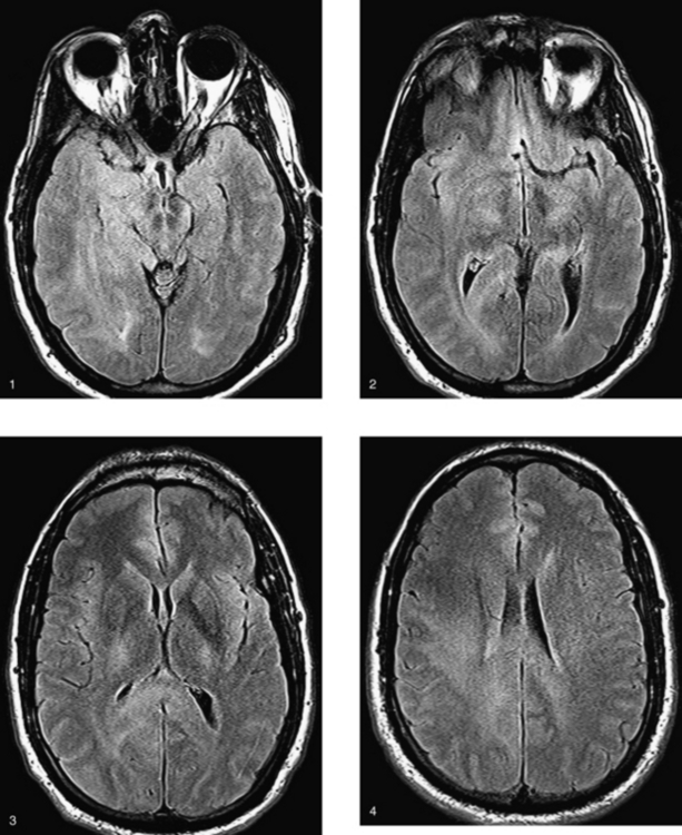
CASE 190 Osmotic Demyelination

Comment

Osmotic demyelination may be seen in underlying systemic processes that have a predilection for electrolyte abnormalities. It is most commonly seen in alcoholics and in chronically debilitated and malnourished patients after rapid correction of hyponatremia. It is not the low serum level but rather the rapidity with which it is corrected that is believed to be responsible for this disorder. Overzealous correction of serum sodium levels may be followed by acute or subacute clinical deterioration, including change in mental status, coma, quadriparesis, extrapyramidal signs, and if unrecognized, death. This process not uncommonly involves extrapontine structures, as in this case. Pathologically, demyelination is noted without a significant inflammatory response, with relative sparing of axons. There is an associated reactive astrocytosis.

When osmotic demyelination is localized only in the pons (central pontine myelinolysis), it is characterized by hyperintensity on T2W images in the central pons, with relative sparing of the peripheral pons. The corticospinal tracts are usually spared. In this case, the first two images are diffusion-weighted images obtained in the acute setting. They show restricted diffusion (hyperintensity) in the regions of acute demyelination in the pons and extrapontine structures involved. The two FLAIR images obtained 1 week later show signal abnormality in the regions of previously noted restricted diffusion (the hyperintensity seen on the diffusion-weighted images resolved). Usually, osmotic demyelination is not associated with enhancement or significant mass effect.

When osmotic demyelination is localized only to the pons, the radiologic diagnosis is usually easy. However, if there is pontine and extrapontine involvement or involvement only in extrapontine structures, the differential diagnosis is somewhat broad, including other demyelinating disorders, encephalitis, and ischemia.

CASE 191 Rosai-Dorfman Disease—Benign Sinus Histiocytosis

Comment

Rosai-Dorfman disease, also known as benign sinus histiocytosis, is a rare disorder in which there is overproduction and accumulation of histiocytes within the lymph nodes. The pathogenesis is unknown, but it may represent an autoimmune disease or a reaction to an infectious agent. Classically, it causes significant painless cervical lymphadenopathy. It may also cause nasal obstruction and enlargement of the palatine tonsils. Extranodal involvement occurs in one-third of cases and may involve the skin, orbit, upper respiratory tract, testes, kidneys, and gastrointestinal tract. The CNS is rarely involved, with approximately 5% of patients with this disease having intracranial manifestations. In approximately two thirds of patients with CNS involvement, the presentation is limited to the brain and spinal cord without lymphadenopathy, as in this case. The typical age of presentation is between 20 and 40 years. Symptoms vary based on the location of involvement.

Intracranial disease may present with headaches, cranial nerve palsies (as in this case), and seizures. More than 90% of patients with CNS disease have involvement of the leptomeninges, with a predominant radiologic presentation of dural-based enhancing masses. It may result in vasogenic edema of the underlying brain parenchyma. On histopathologic evaluation, Rosai-Dorfman disease of the meninges is similar to that in the lymph nodes. The dura is thickened and fibrotic, resulting in its hypointensity on T2W, and it contains inflammatory cells, with a predominance of lymphocytes and plasma cells. Interspersed are pale histiocytes with vacuolated cytoplasm. The differential diagnosis includes granulomatous disease, such as sarcoid, lymphoma (Hodgkin’s disease), and Langerhans’ histiocytosis. When the mass is isolated, it may closely resemble a meningioma.

Meningeal Rosai-Dorfman disease is primarily treated with surgical resection. Disease progression after surgery is reportedly uncommon. Progressive disease occurs most often when there is multiple-organ involvement. In such cases, as well as in cases like this one, where there is diffuse meningeal involvement, other therapies, including steroids, vincristine, cyclophosphamide (Cytoxan), and radiation, have had variable success.

CASE 192 Cystic Necrotic Brain Mass—Part I

Comment

The differential diagnosis of a single ring-enhancing lesion with prominent associated vasogenic edema includes metastatic disease, a brain abscess, and a high-grade primary glial neoplasm. Less common causes of large ring-enhancing lesions include demyelination and resolving hematoma. Advanced MR imaging techniques, such as diffusion-weighted imaging, spectroscopy, and perfusion imaging, can sometimes be helpful in distinguishing among these. Abscesses may show cytosolic amino acids, including alanine and valine, as well as other metabolites, such as lactate and pyruvate, which are end-products of bacterial metabolism. However, some necrotic tumors may have spectra with many of these features (lactate, amino acids).

Diffusion-weighted MR imaging provides information about the diffusional properties of water in the brain related to the local cellular microenvironment. The central cavity of a brain abscess frequently has restricted diffusion (restricted mobility of water protons) as a result of several factors, including, but not limited to, the cellularity and viscosity of pus, as well as the presence of large molecules, such as fibrinogen. It is important to recognize, however, that abscesses evolve over time on treatment, and so do their imaging characteristics. Changes in their signal characteristics on diffusion-weighted imaging are related to increases in free water and associated changes in the viscosity of the fluid, different bacteria, and the concentration of inflammatory cells.

CASE 193 Cystic Necrotic Brain Mass—Part II—Metastatic Adenocarcinoma of the Lung

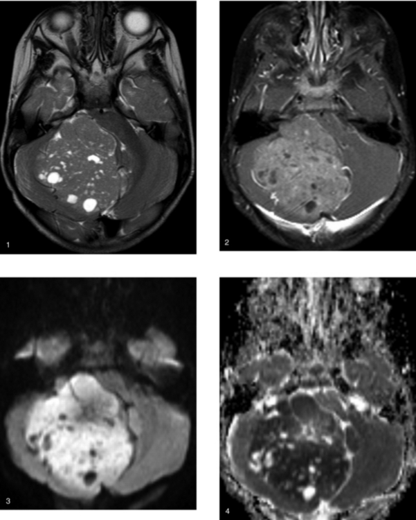
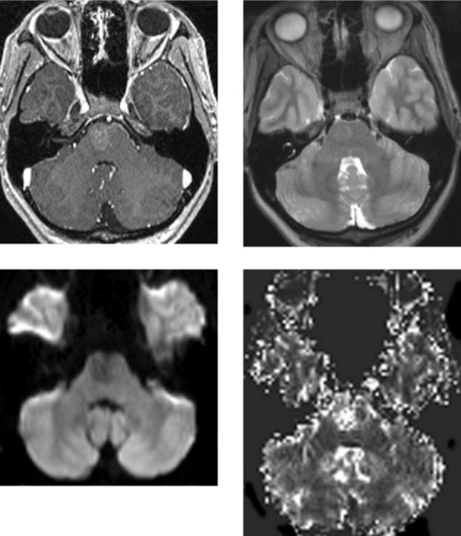
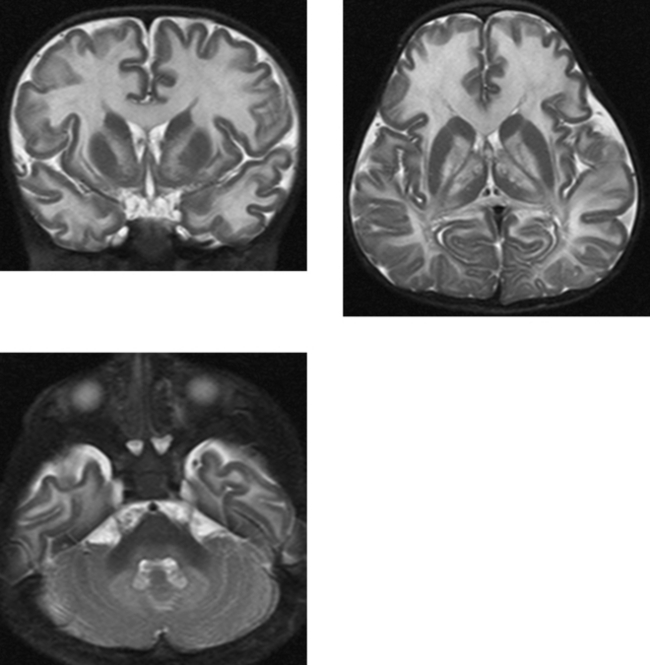
CASE 194 Rhombencephalosynapsis

Comment

Rhombencephalosynapsis is an anomaly of the cerebellar vermis that is agenetic or hypoplastic. There is fusion of the cerebellar hemispheres, with variable fusion of other posterior fossa structures, including the middle cerebellar peduncles, cerebellar dentate nuclei, and superior and inferior colliculi. In the cerebellar hemispheres, the orientation of the folia is disorganized. They are usually transverse in configuration, extending across the midline without intervening vermis. MR imaging typically shows an absent or severely hypoplastic vermis, with fusion of the cerebellar hemispheres. There is usually posterior pointing of the fourth ventricle. Associated supratentorial anomalies include partial or complete absence of the septum pellucidum, a hypoplastic anterior commissure, ex vacuo enlargement of the ventricular system related to surrounding volume loss of the brain parenchyma, and fusion of the thalami. Hypertelorism and migrational anomalies have also been reported with this condition. The clinical presentation is more commonly related to the associated supratentorial anomalies.

Joubert’s syndrome, another dysplasia of the posterior fossa contents, is characterized by severe hypoplasia or aplasia of the cerebellar vermis. It has a characteristic imaging appearance, including a “bat-wing” configuration of the fourth ventricle, as well as a horizontal orientation of the superior cerebellar peduncles. Unlike rhombencephalosynapsis, the cerebellar hemispheres are apposed in the midline, but are not fused. Associated supratentorial anomalies are uncommon.

CASE 195 Progressive Multifocal Leukoencephalopathy

Comment

Before the AIDS epidemic, PML was largely seen in a spectrum of immunocompromised patients, including those with hematologic malignancies (leukemia and lymphoma), those who had undergone organ transplantation, patients taking immunosuppressive drugs, and those with autoimmune disorders. However, over the last few decades, the majority of cases of PML have been noted in patients with HIV infection. Progressive multifocal leukoencephalopathy is caused by infection of the oligodendrocytes with a papovavirus (JC virus). Histologically, multifocal regions of demyelination involve the subcortical U-fibers.

The clinical presentation of PML includes focal neurologic deficits (hemiparesis), visual symptoms, and especially progressive cognitive decline. The infection is rapidly progressive, with continued neurologic decline, CNS demyelination, and death usually occurring within 6 months to a year from the onset of symptoms.

MR imaging is far more sensitive than CT in defining the number and extent of lesions in PML. On CT, PML usually appears as focal regions of hypodensity within the white matter, usually without mass effect or enhancement. On MR imaging, increased T2W signal intensity with associated T1W hypointensity is noted in the involved white matter. PML has a predilection to involve the subcortical white matter, although the deep white matter is also commonly involved. There is a slight preference for involvement of the parietal and occipital white matter, but any area of the brain may be affected, including the cerebellum, as in this case. Although single focal lesions may be seen, multifocal lesions typically occur and may or may not be bilaterally symmetric in distribution. Unilateral multifocal distribution may also occur. Mass effect or enhancement is less common in PML, occurring in 5% to 10% of cases.

CASE 196 Sphenoid Dysplasia and Brain Dysplasia—Neurofibromatosis Type 1 (von Recklinghausen’s Disease)

Comment

There is still some controversy over what the hyperintense lesions on T2W imaging represent in patients with neurofibromatosis type 1. These lesions are frequently located in the peduncles or deep gray matter of the cerebellum, the basal ganglia, the white matter of the cerebral hemispheres, and the brainstem, especially the pons. Some of the lesions may be mildly hyperintense on unenhanced T1W imaging. In this case, such lesions are seen in the right cerebellum and medial left temporal lobe on images obtained in 1999. On a follow-up study in 2007, the cerebellar lesion has resolved and the left temporal lobe lesion is smaller. Heterotopias and hamartomas have been suggested as the cause of these T2W hyperintense foci. Heterotopias are related to anomalous migration of gray matter, whereas hamartomas represent nonneoplastic proliferation of normal brain tissue in abnormal locations. The pathologic literature has described gliosis and vacuolar and spongiotic change. Myelin vacuolization in areas of dysplastic white matter seems to be commonly suggested. These lesions typically appear during the first decade of life, and may regress over time. If there is growth or enhancement of one of these foci, the possibility of an astrocytoma must be considered. Astrocytomas are seen with an increased incidence in neurofibromatosis type 1. It is believed by many (but not all) that these are separate lesions and that they do not arise from regions of dysplastic brain.

The primary manifestation of mesodermal dysplasia is hypoplasia or absence of the greater wing of the sphenoid bone. This may present with encephaloceles, as in this case, or with pulsatile exophthalmos. Buphthalmos is seen with increased frequency in neurofibromatosis type 1, and it refers to an enlarged globe caused by increased intraocular pressure secondary to obstruction of the canal of Schlemm from a mass or membranes composed of aberrant mesodermal tissue.

Neurofibromatosis type 1 can be diagnosed when two or more of the following criteria are present: a first-degree relative with neurofibromatosis type 1, one plexiform neurofibroma or two or more neurofibromas of any type, six or more café-au-lait spots, two or more Lisch nodules (iris hamartomas), axillary or inguinal freckling, optic pathway glioma, or a characteristic bone abnormality (dysplasia of the greater wing of the sphenoid, overgrowth of a digit or limb, pseudarthrosis, lateral thoracic meningocele, dural ectasia with vertebral dysplasia).

CASE 197 Panhypopituitarism with Absence of the Infundibulum
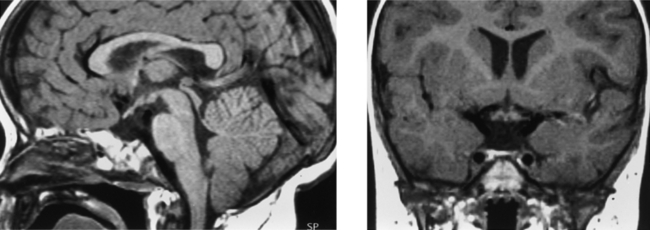
Comment

This case shows an absent or diminutive anterior pituitary lobe, absence of the pituitary stalk, and absence of the normal posterior pituitary “bright” spot in the pituitary sella. The normal neurohypophysis is of high signal intensity on unenhanced T1W images; however, the exact etiology of this is debated, with explanations including vasopressin, phospholipid, and neurophysin stored in neurosecretory granules of the posterior pituitary gland. High signal intensity at the apex or median eminence of the pituitary stalk is characteristic of an ectopic posterior pituitary gland. The differential diagnosis of high signal intensity along the apex of the stalk at the floor of the third ventricle includes a tuber cinereum lipoma or fat in the marrow of the tip of the dorsum sella. In the latter two conditions, the normal posterior pituitary “bright” spot in the sella is present. Disappearance of the “hyperintensity” on fat-suppressed imaging could also confirm the presence of fat (lipoma or marrow). Absence of the normal posterior pituitary high signal intensity may be seen in Langerhans’ cell histiocytosis and hemosiderosis.

The neurohypophysis is ectopic in a subset of patients with pituitary dwarfism. Specifically, low growth hormone levels associated with other pituitary hormonal deficiencies may be accompanied by an ectopic posterior pituitary gland and absence of the infundibulum on MR imaging. Failure of development of the infundibulum and posterior pituitary gland may be an isolated condition or may be part of a larger brain anomaly. It is believed that an ectopic posterior pituitary gland is related to an injury to the pituitary infundibulum, which normally transmits hormonal mediators from the hypothalamus to the neurohypophysis through a rich venous plexus. An ectopic posterior pituitary gland has also been associated with traumatic transection of the pituitary stalk.

CASE 198 Progressive External Ophthalmoplegia Syndrome—due to Mitochondrial Defect

Comment

This patient presented with external ophthalmoplegia syndrome that was progressive over a 20-year period. The patient also had dysphagia and vertigo. Chronic progressive external ophthalmoplegia (CPEO) is a disorder characterized by slowly progressive paralysis of the extraocular muscles. It is usually bilateral and symmetrical. The ciliary and iris muscles are not involved. CPEO is the most frequent manifestation of mitochondrial myopathies. CPEO in association with mutations in mitochondrial DNA may occur in the absence of any other clinical sign, but usually, it is associated with skeletal muscle weakness. This patient’s extensive evaluation was notable for a mutation in a region of a nuclear-encoded gene that encodes a DNA polymerase involved in mitochondrial gene maintenance. The mutation is in a gene that has been implicated in these syndromes, and it is in a region where pathogenic mutations have been identified. The region is highly conserved from fruit flies to humans. All of these factors make it likely to be a pathogenic mutation. These mutations are usually associated with DNA depletion syndromes, in which there is less DNA overall in the mitochondria. The treatment is limited and involves a “mitochondrial cocktail”: vitamin C, vitamin E, coenzyme Q10, creatine, carnitine, riboflavin, pantothenate, thiamine, and lipoic acid.

The differential diagnosis includes other progressive external ophthalmoplegia syndromes, such as SANDO and Kearns-Sayre, as well as ocular pharyngeal dystrophy and myasthenia gravis. Kearns-Sayre syndrome is a mitochondrial myopathy in which the onset of CPEO occurs before 20 years of age and pigmentary retinopathy occurs as well. Kearns-Sayre syndrome also is associated with cardiac conduction defects, elevated cerebrospinal fluid protein, and a cerebellar syndrome.

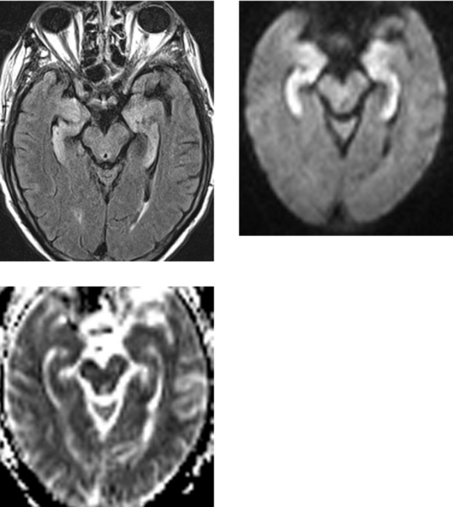
CASE 199 Limbic Encephalitis—Paraneoplastic Syndrome (Small Cell Lung Carcinoma)
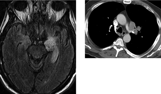
Comment

Paraneoplastic syndromes affecting the CNS represent a spectrum of neurologic manifestations that are associated with extracranial cancers, but are not the result of direct invasion of the CNS by tumor. They occur in fewer than 1% of patients with cancer; however, in 33% to 50% of patients with a paraneoplastic syndrome, the syndrome develops before the diagnosis of systemic neoplasm. Such syndromes include limbic encephalopathy, cerebellar degeneration, opsoclonus or myoclonus, retinal degeneration, Lambert-Eaton myasthenic syndrome, and myelopathy. Lung cancer, particularly small cell carcinoma, is the most common malignancy associated with neurologic paraneoplastic syndromes. However, these syndromes may be seen in ovarian carcinoma, testicular germ cell tumors, gastrointestinal cancer, Hodgkin’s disease, breast cancer, and neuroblastoma in children. The cause of paraneoplastic syndromes is unknown; however, the most widely accepted theory is that they occur as a result of an autoimmune disorder. Circulating autoantibodies have been identified in several paraneoplastic syndromes. Anti-Yo is specific to paraneoplastic cerebellar degeneration associated with breast and ovarian cancer, and anti-Hu is most often associated with paraneoplastic limbic encephalitis.

Paraneoplastic limbic encephalitis may present with a change in mental status, personality changes, and memory impairment. CT may be unremarkable. On MR imaging, high signal intensity may be identified in the medial temporal lobes on T2W and FLAIR images. Mild enhancement may occur. Involvement of the hypothalamus may also be noted. On pathologic evaluation, nonspecific inflammatory changes and cellular infiltrates are identified without the presence of tumor or viral inclusions. Treatment of the primary malignancy may result in improvement of the neurologic symptoms.

CASE 200 von Hippel-Lindau Disease

Comment

This case shows a cystic mass with a solid enhancing mural nodule in the region of the area postrema of the medulla, as well as multiple small enhancing cerebellar lesions. Also noted is one of many superficial enhancing nodules along the pial surface of the spinal cord. Because this patient is an adult and the lesions are multiple, juvenile pilocytic astrocytoma is not a consideration. The differential diagnosis includes multiple hemangioblastomas seen in von Hippel-Lindau disease and, less likely, metastatic disease.

Although von Hippel-Lindau disease is a neurocutaneous syndrome, neurocutaneous is a misnomer in that there are no cutaneous manifestations. The hallmark lesion is the hemangioblastoma. Approximately 20% of patients with hemangioblastomas have von Hippel-Lindau disease. Conversely, up to 45% of patients with von Hippel-Lindau disease have CNS hemangioblastomas. In von Hippel-Lindau disease, these neoplasms are multiple in at least 40% of cases. Hemangioblastomas occur most commonly in the cerebellum and retina, although they may also arise in the brainstem (especially the medulla), the spinal cord, and (rarely) the cerebrum and viscera outside of the CNS. In addition to CNS lesions, patients with von Hippel-Lindau disease also have a spectrum of findings in other organs. Cysts in the pancreas occur in the majority of patients, but cysts may also be seen in the kidneys, liver, and epididymis in men and ovaries in women. Renal cell carcinomas are common and are different from garden variety renal cell adenocarcinoma in that in von Hippel-Lindau disease they are frequently bilateral, small, and less malignant. There is an approximately tenfold increase in the incidence of pheochromocytomas in these patients. Polycythemia related to the production of erythropoietin by the hemangioblastomas is not uncommon. Diagnostic criteria for von Hippel-Lindau disease include more than one CNS or retinal hemangioblastoma; a CNS hemangioblastoma and at least one visceral abnormality; and a family history of von Hippel-Lindau disease and at least one of the visceral manifestations.