• Endemic in Central America, Brazil, northern Argentina, Venezuela
Affects millions of patients in these countries
• Reported in southern USA
(Left) Esophagram in a 56-year-old woman with known Chagas disease (who had recently complained of dysphagia) shows decreased esophageal motility with mild narrowing at the gastroesophageal junction . The esophagus is only mildly dilated.
(Right) Esophagram shows esophageal dilatation. Esophageal contractions were seen during fluoroscopy, but there was failure of the lower esophageal sphincter to relax.
(Left) Esophagram on a 48-year-old man from Brazil with known Chagas disease shows esophageal dilatation with abrupt tapering at the gastroesophageal junction and lack of peristalsis. The degree of esophageal involvement in Chagas disease is quite variable, ranging from normal to megaesophagus.
(Right) PA radiograph shows global cardiomegaly and clear lung fields in a patient with chronic cardiomyopathy from Chagas disease. There is no evidence of pulmonary congestion or pleural effusion.
Hematoxylin and eosin stain from a cardiac biopsy shows extensive infiltration of the myocardium with inflammatory cells . The myocardial cells are destroyed in the central area of the photomicrograph and Trypanosoma cruzi , the causative parasite, is seen.


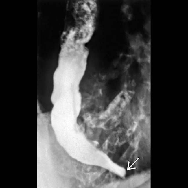
 . The esophagus is only mildly dilated.
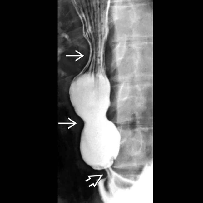
 were seen during fluoroscopy, but there was failure of the lower esophageal sphincter
  to relax.
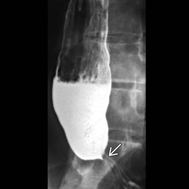
 and lack of peristalsis. The degree of esophageal involvement in Chagas disease is quite variable, ranging from normal to megaesophagus.
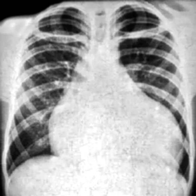
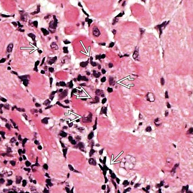
 . The myocardial cells are destroyed in the central area of the photomicrograph and Trypanosoma cruzi
  , the causative parasite, is seen.

