Chapter 17 Cervical Plexus
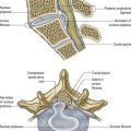
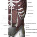
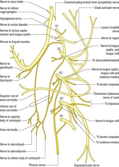
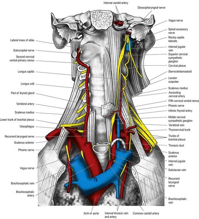
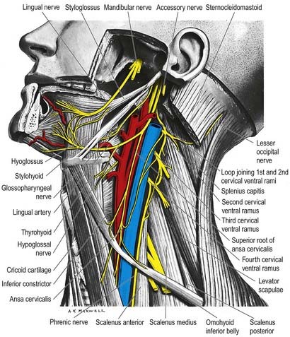
The cervical plexus is formed by the ventral rami of the upper four cervical nerves, and it supplies some neck muscles, the diaphragm and areas of skin on the head, neck and chest (Figs 17.1–17.3; see also Fig. 11.10). It is situated in the neck opposite a line drawn down the side of the neck from the root of the auricle to the level of the upper border of the thyroid cartilage. It is deep to the internal jugular vein, the deep fascia and sternocleidomastoid, and anterior to scalenus medius and levator scapulae. Each ramus, except the first, divides into ascending and descending parts that unite in communicating loops. From the first loop (C2 and C3), superficial branches supply the head and neck; cutaneous nerves of the shoulder and chest arise from the second loop (C3 and C4). Muscular and communicating branches arise from the same nerves. The branches are superficial or deep. The superficial branches perforate the cervical fascia to supply the skin, whereas the deep branches generally supply the muscles. The superficial branches either ascend (lesser occipital, great auricular and transverse cutaneous nerves) or descend (supraclavicular nerves). The deep branches form medial and lateral series.
Deep Branches—Medial Series
Communicating Branches
Communicating branches pass from the loop between the first and second cervical rami to the vagus and hypoglossal nerves and to the sympathetic trunk. The hypoglossal branch later leaves the hypoglossal nerve as a series of branches—namely, the meningeal, superior root of ansa cervicalis and nerves to thyrohyoid and geniohyoid. A branch also connects the fourth and fifth cervical rami. The first four cervical ventral rami each receive a grey ramus communicans from the superior cervical sympathetic ganglion.
The superior root of the ansa cervicalis (descendens hypoglossi; see Fig. 17.1) leaves the hypoglossal nerve where it curves around the occipital artery and then descends anterior to or in the carotid sheath. It contains only fibres from the first cervical spinal nerve. After giving a branch to the superior belly of omohyoid, it is joined by the inferior root of the ansa from the second and third cervical spinal nerves. The two roots form the ansa cervicalis (ansa hypoglossi), from which branches supply sternohyoid, sternothyroid and the inferior belly of omohyoid. Another branch descends anterior to the vessels into the thorax to join the cardiac and phrenic nerves.
Muscular Branches
Inferior Root of Ansa Cervicalis
The inferior root of the ansa cervicalis (nervus descendens cervicalis) is formed by the union of branches from the second and third cervical rami (see Fig. 17.1). It descends on the lateral side of the internal jugular vein, crosses it a little below the middle of the neck and continues forward to join the superior root anterior to the common carotid artery, forming the ansa cervicalis (ansa hypoglossi), from which all infrahyoid muscles except the thyrohyoid are supplied. The inferior root comes from the second and third cervical ventral rami in approximately 75% cases, from the second to fourth in 15% and from the third alone in 5%. Occasionally, it may be derived either from the second alone or from the first to third.
Phrenic Nerve
The phrenic nerve arises chiefly from the fourth cervical ventral ramus, but it also has contributions from the third and fifth. It is formed at the upper part of the lateral border of scalenus anterior and descends almost vertically across its anterior surface behind the prevertebral fascia. It descends posterior to the sternocleidomastoid, the inferior belly of omohyoid (near its intermediate tendon), the internal jugular vein, transverse cervical and suprascapular arteries and, on the left, the thoracic duct. At the root of the neck it runs anterior to the second part of the subclavian artery, from which it is separated by the scalenus anterior (according to some accounts, on the left side the nerve passes anterior to the first part of the subclavian artery), and posterior to the subclavian vein. The phrenic nerve enters the thorax by crossing medially in front of the internal thoracic artery.