[level-membership-for-basic-science-category]65
Cerebrospinal and other body fluids
Cerebrospinal fluid
Xanthochromia
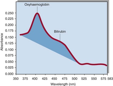
Xanthochromia simply means yellow discoloration of the CSF. It results from the presence of bilirubin derived from red blood cells (RBCs) that have undergone in vivo lysis. In vitro lysis of RBCs, e.g. a traumatic LP, produces only oxyhaemoglobin, and not bilirubin. It can be detected by visual inspection, but this is unreliable, and where possible scanning spectrophotometry should be used instead. This involves measuring the absorbance of the CSF specimen across a range of wavelengths; the blood pigments have characteristic absorbance peaks (Fig 65.1).
Meningitis
Meningitis refers to inflammation of the meninges which line the central nervous system (CNS). Bacterial meningitis presents acutely and is a medical emergency. CSF biochemistry tends to reflect the nature of the infective organism (Table 65.1) but is characteristic rather than diagnostic. Microbiological analysis should take priority. It is important when interpreting the relative concentrations of, for example, glucose in the CSF to take a blood sample for comparison.
Table 65.1
CSF parameters in health and some common disorders
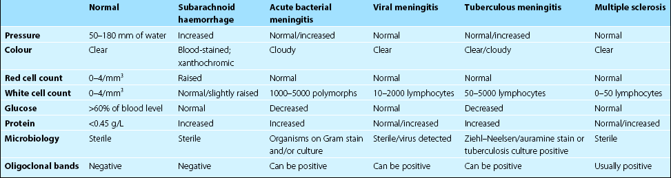
From Haslett C et al, Davidson’s Principles and Practice of Medicine. Churchill Livingstone, Edinburgh, 2002.
Other conditions
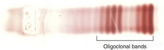
CSF electrophoresis may also reveal the presence of oligoclonal bands (Fig 65.2). If these are not seen also in the serum, they reflect local (i.e. CNS) synthesis of immunoglobulin. Ninety per cent of patients with multiple sclerosis (MS) have these bands, but they are not specific for this condition. Thus their absence in cases of suspected MS is more diagnostically useful than their presence.
Identification of body fluids
Other fluids
 Clinical note


[/level-membership-for-basic-science-category][not-level-membership-for-basic-science-category]65
Cerebrospinal and other body fluids
Cerebrospinal fluid
Xanthochromia
Xanthochromia simply means yellow discoloration of the CSF. It results from the presence of bilirubin derived from red blood cells (RBCs) that have undergone in vivo lysis. In vitro lysis of RBCs, e.g. a traumatic LP, produces only oxyhaemoglobin, and not bilirubin. It can be detected by visual inspection, but this is unreliable, and where possible scanning spectrophotometry should be used instead. This involves measuring the absorbance of the CSF specimen across a range of wavelengths; the blood pigments have characteristic absorbance peaks (Fig 65.1).
Meningitis
Meningitis refers to inflammation of the meninges which line the central nervous system (CNS). Bacterial meningitis presents acutely and is a medical emergency. CSF biochemistry tends to reflect the nature of the infective organism (Table 65.1
[/not-level-membership-for-basic-science-category]
